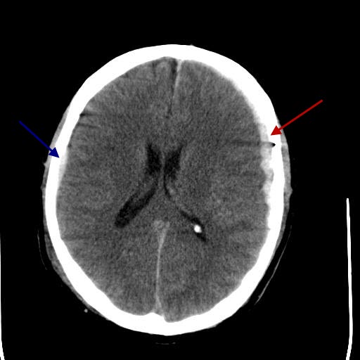
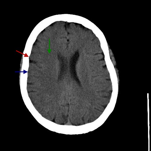
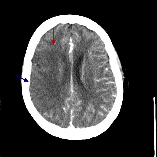
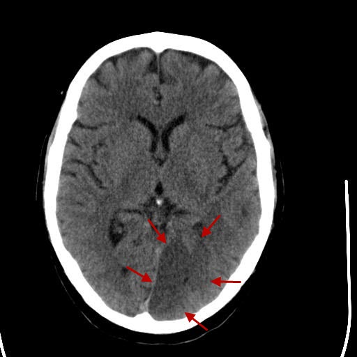
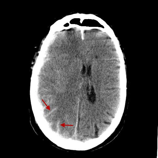
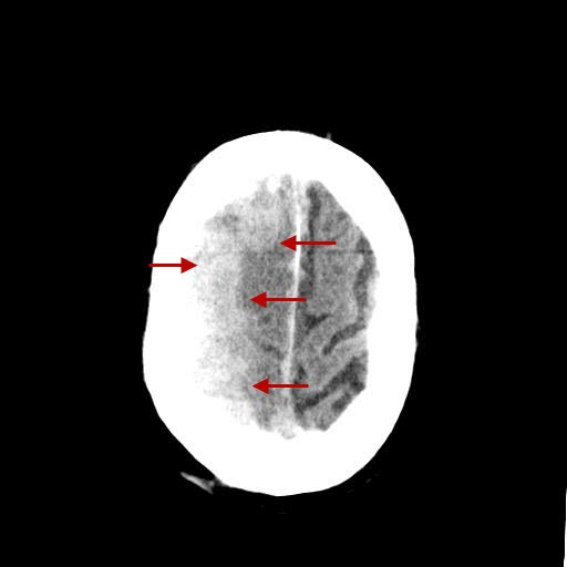
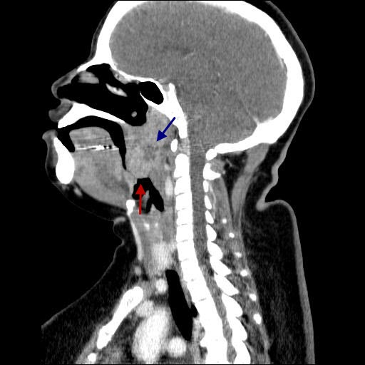
NeuroHead - Acute right MCA stroke Head - Left PCA infarct Head - Subarachnoid hemorrhage, subdural bleed, brain edema, cerebral vasospasm Neck - Tonsillar abscess Neuro: HeadDiagnosis: Bilateral subdural hematomas and parenchymal bleed (CT)There is a focal extra axial hematoma overlying portions of the left frontal, parietal and temporal lobes compatible with a subdural hematoma. Subdural hematomas cross sutures - epidural hematomas don't. Did you also see the additional extra axial hematoma overlying the right posterior frontal/parietal lobe measuring approximately 3 mm in greatest diameter?Neuro: HeadDiagnosis: Acute right MCA stroke (CTA, CT)Non contrast head CT shows in the right frontal lobe loss of corticomedullary (gray-white matter) differentiation, also known as ribbon sign. There is also mild associated parenchymal swelling. Contrast enhanced head CT (CTA) demonstrates absent perfusion involving the right MCA territory. This appears to be larger than 1/3 of the MCA territory, which does not make it amendable anymore for lysis therapy. Image stack 1 are MIPs, second stack time to peak imagesNeuro: HeadDiagnosis: Left PCA infarct (CT)A well-defined wedge-shaped hypodensity is present in the left occipital lobe medially extending to occipital pole representing acute ischemic infarct in territory of left PCA, with involvement of primary visual cortex.Neuro: HeadDiagnosis: Subarachnoid hemorrhage, subdural bleed, brain edema, cerebral vasospasm (CT, CTA)Extensive subarachnoid hemorrhage, subdural bleed, brain edema, cerebral vasospasm. There is blood in the subarachnoid space, notable as hyperdense material within the sulci. A subdural hematoma is also noted along the right hemisphere. CTA and volume rendered images demonstrate diminuitive intracerebral vessels, indicating vasospasm.Neuro: NeckDiagnosis: Tonsillar abscess (CT)There is an about 4.4 x 3.1 x 3.6 cm tonsillar soft tissue swelling, with a 1.5 x 1.1 x 1.4 cm central area of low density on the right side, likely a tonsillar abscess. Note also the narrowing of the naso- and oropharynx (red arrow in image 2 and 3) due to extensive soft tissue swelling.Home :: Contact Us Make On Call Radiology your homepage :: Add On Call Radiology to your favorites |