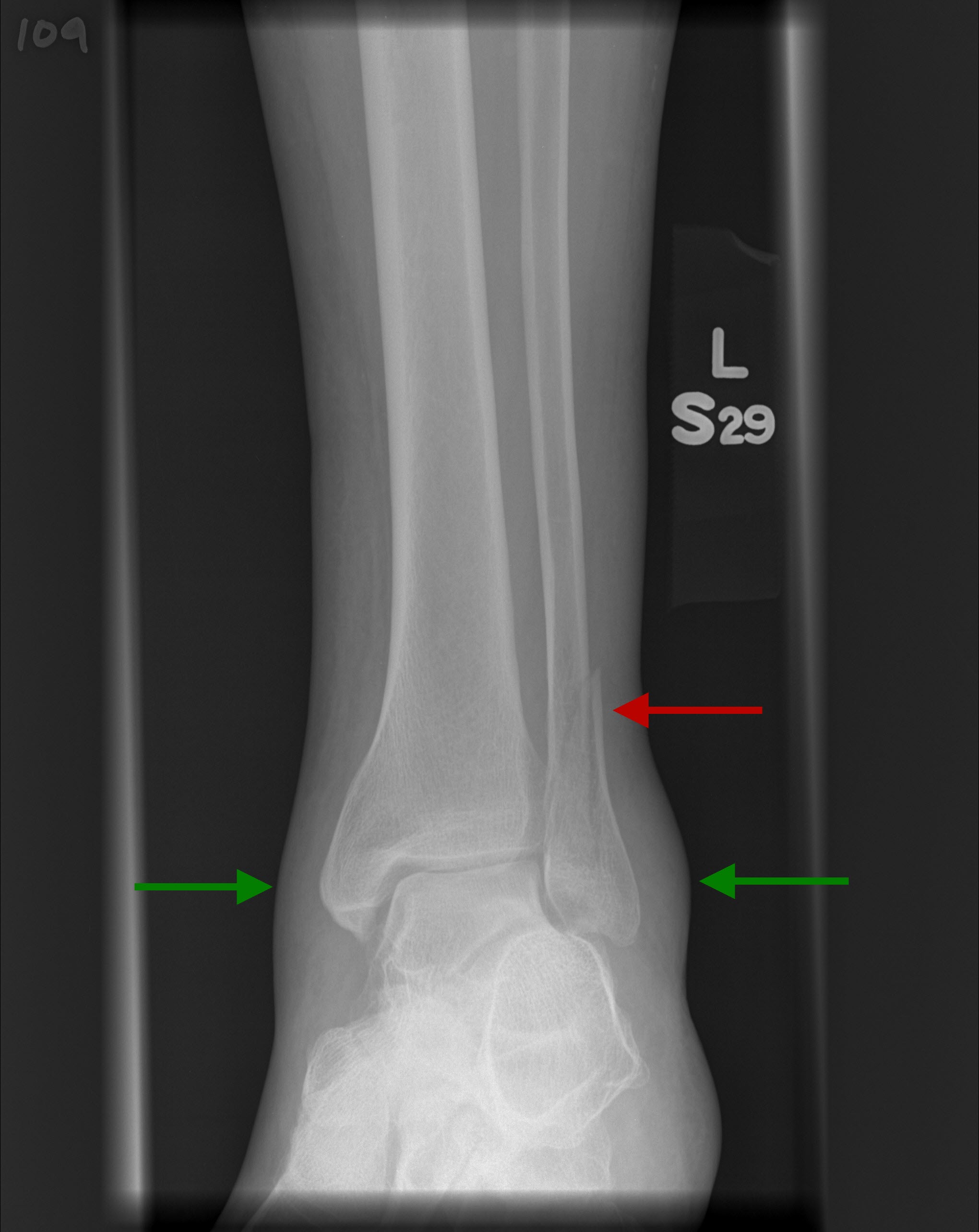
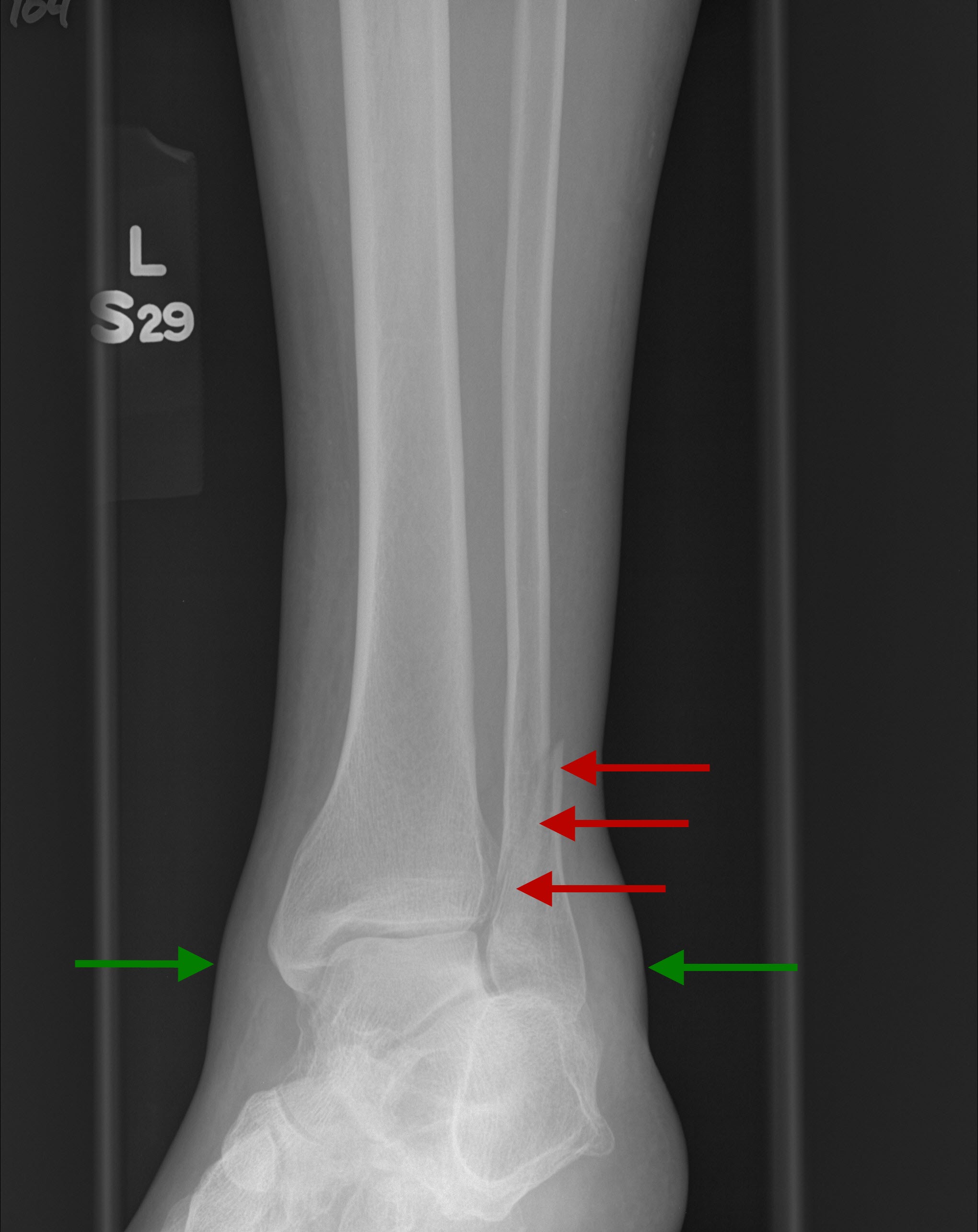
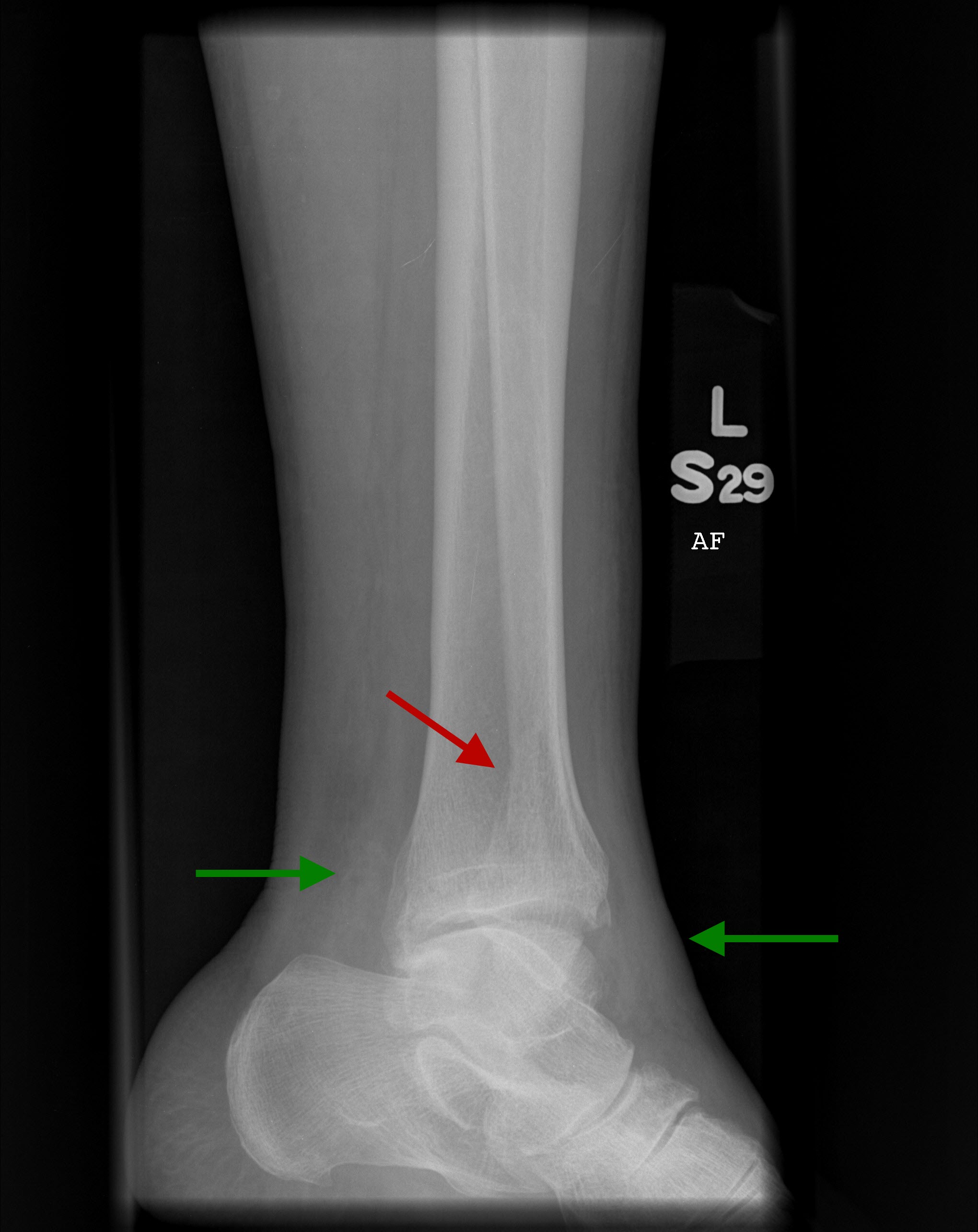
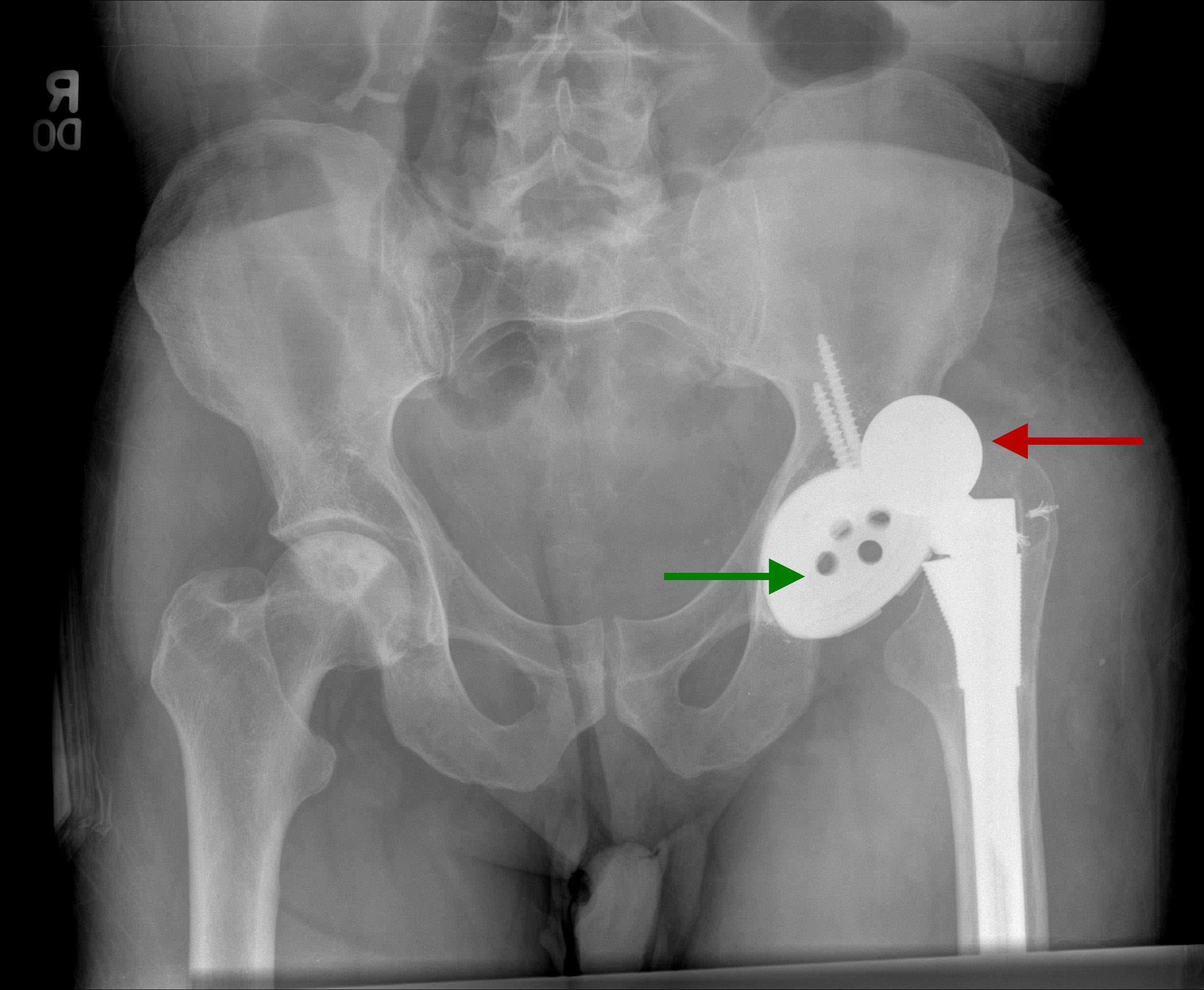
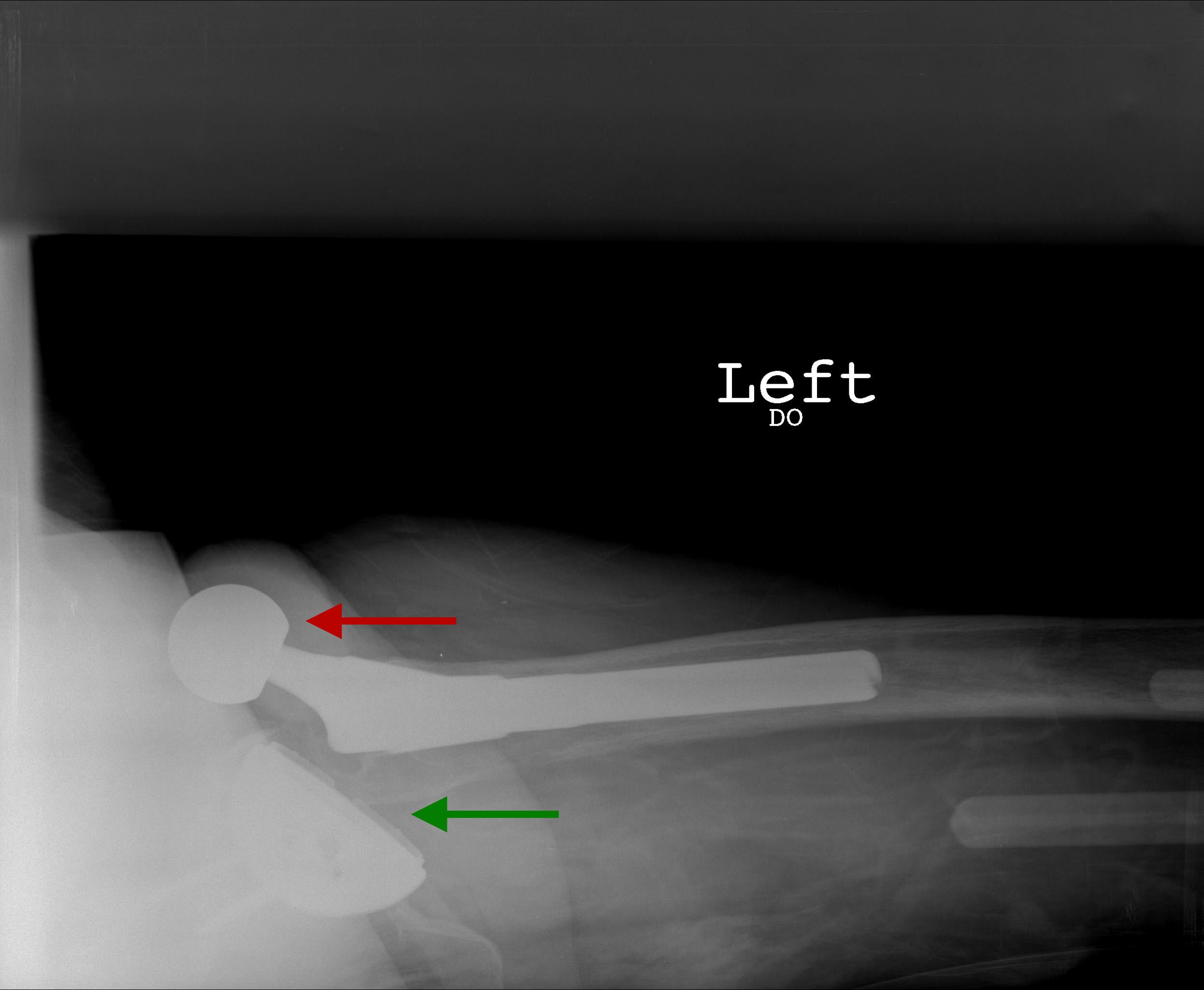
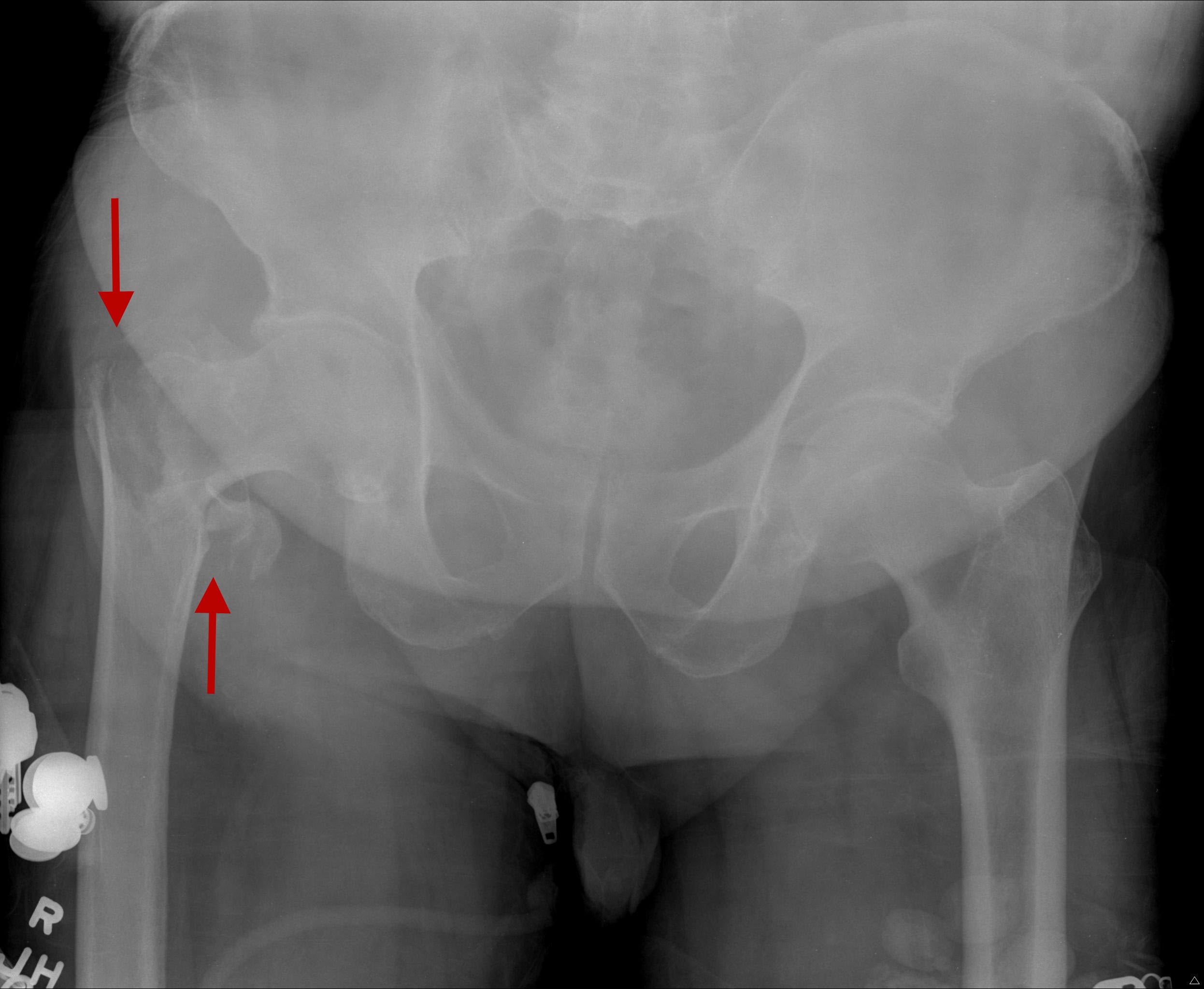
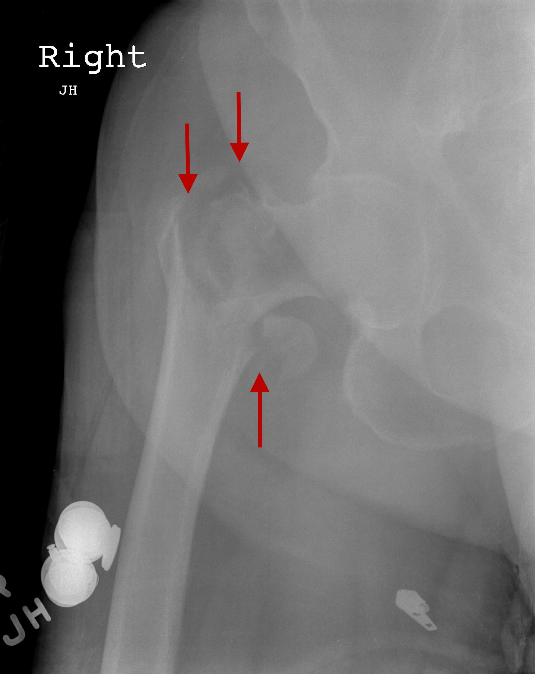
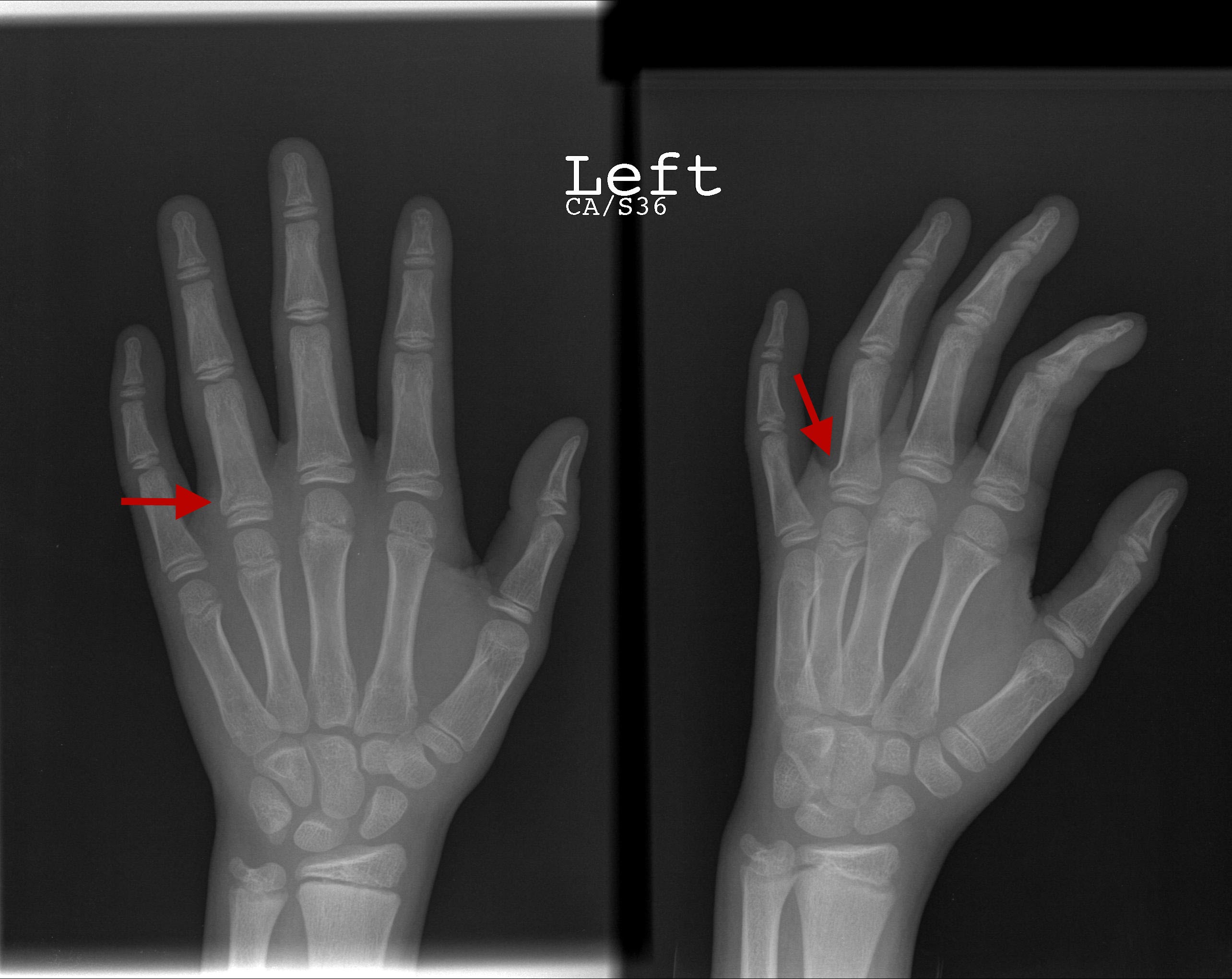
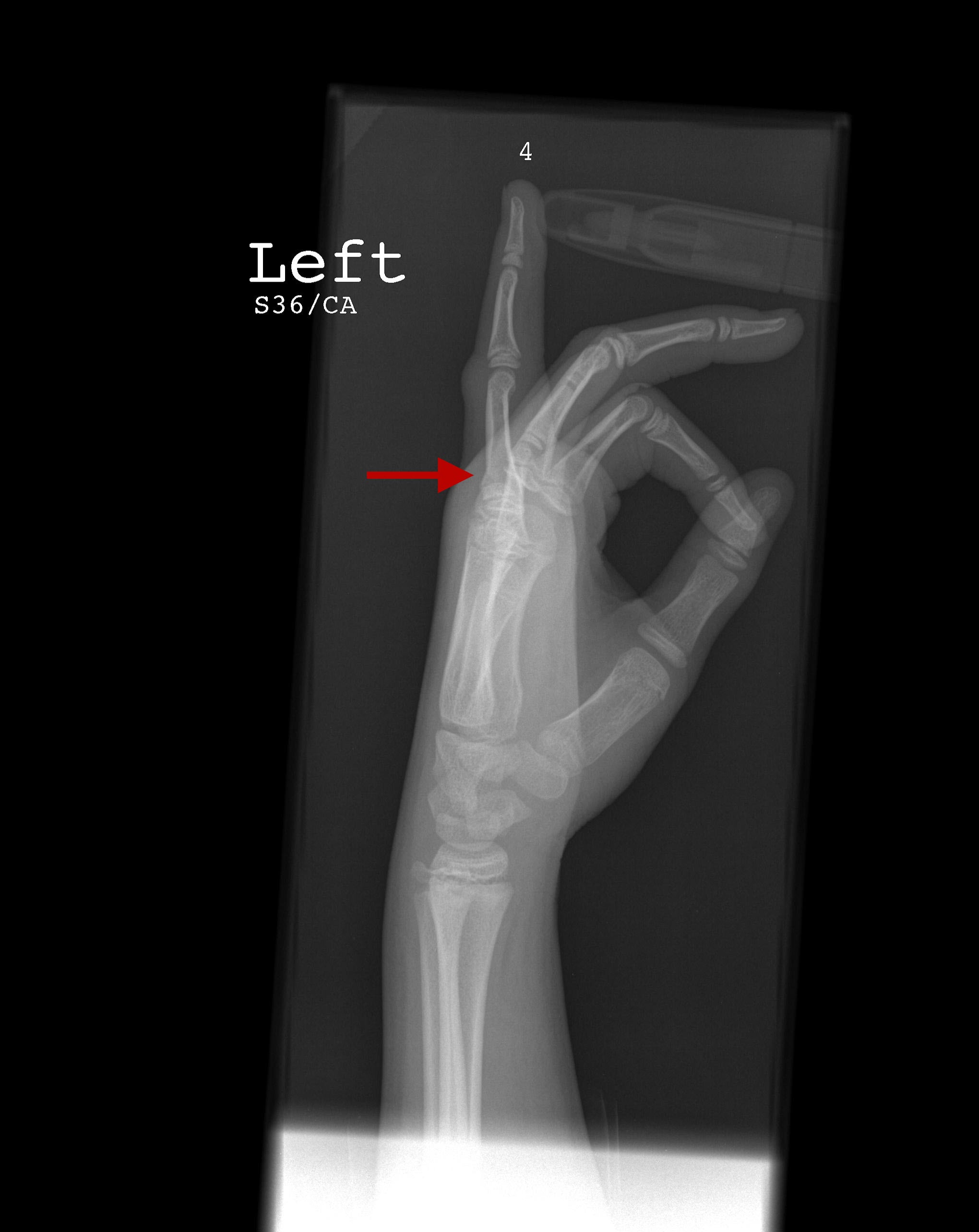
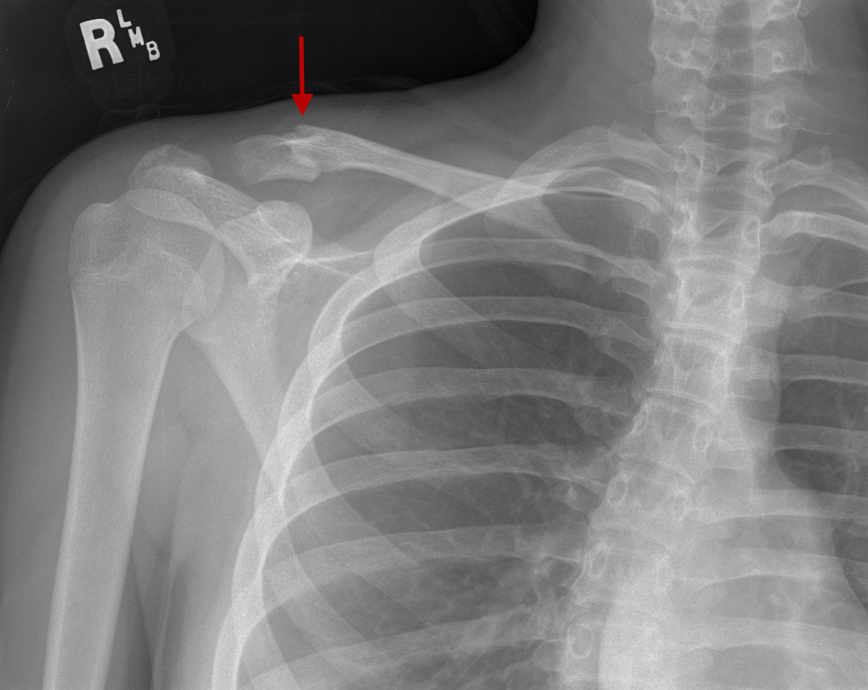
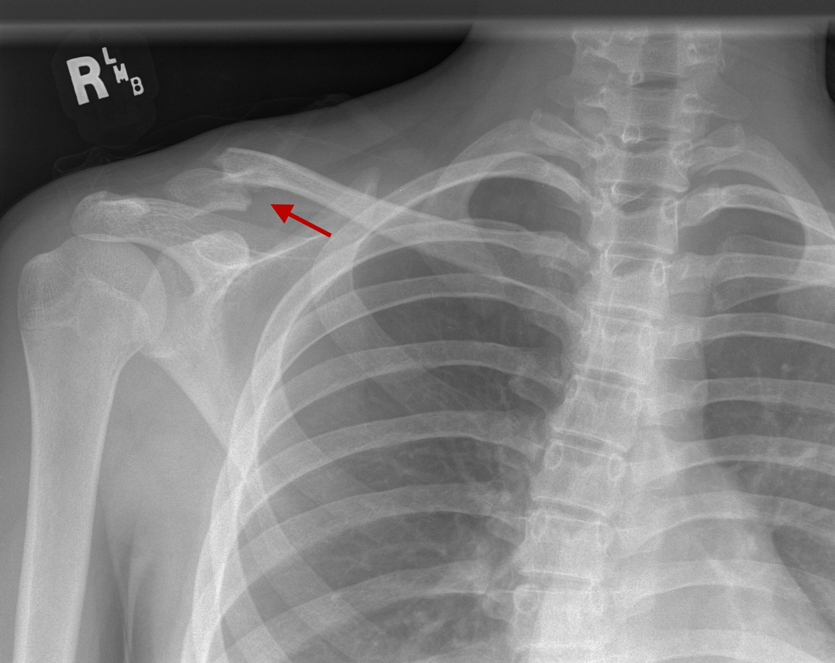
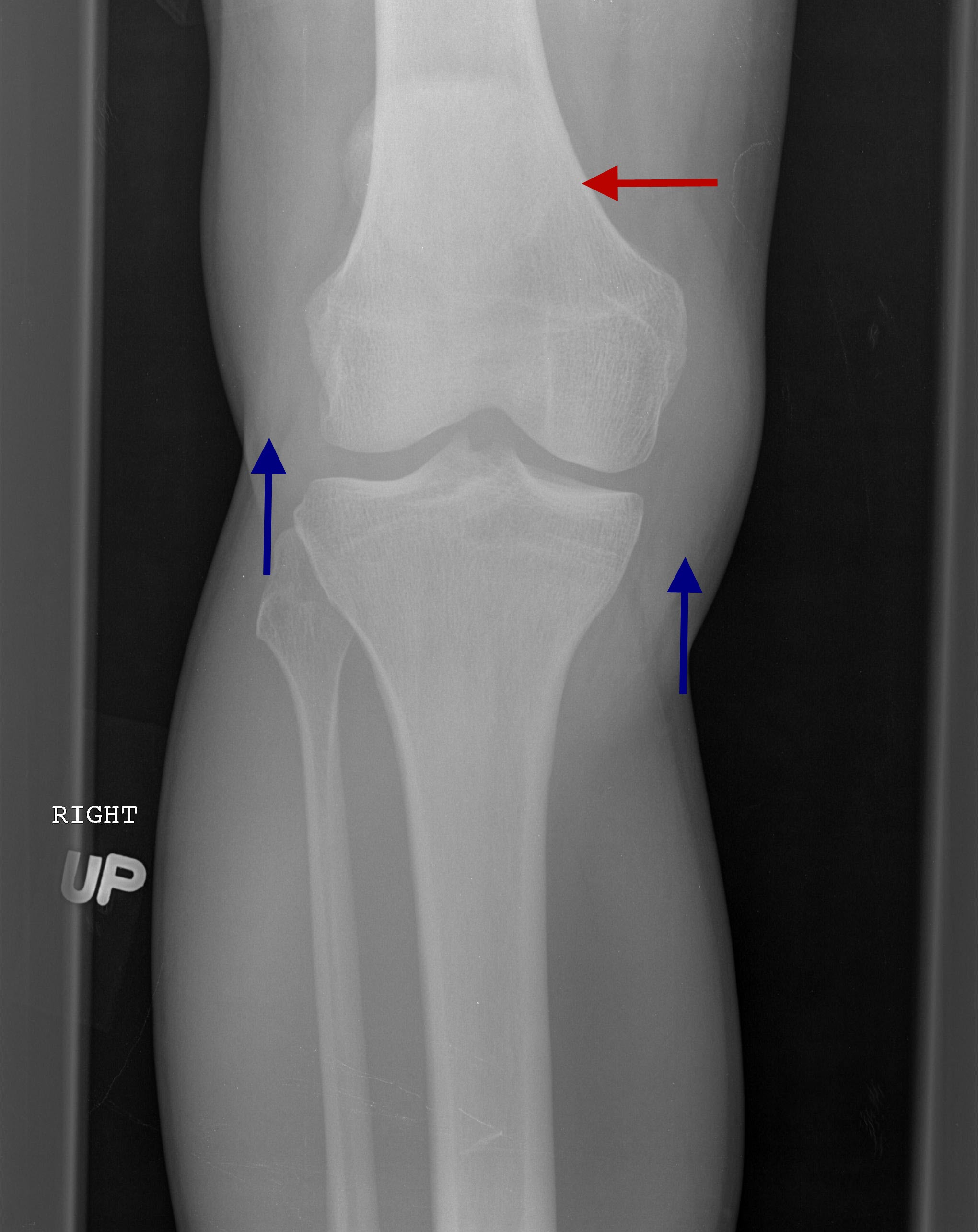
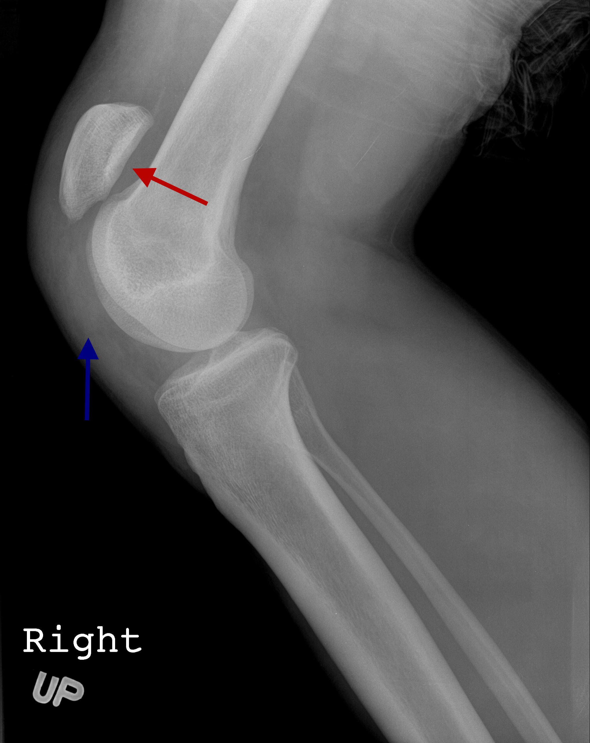
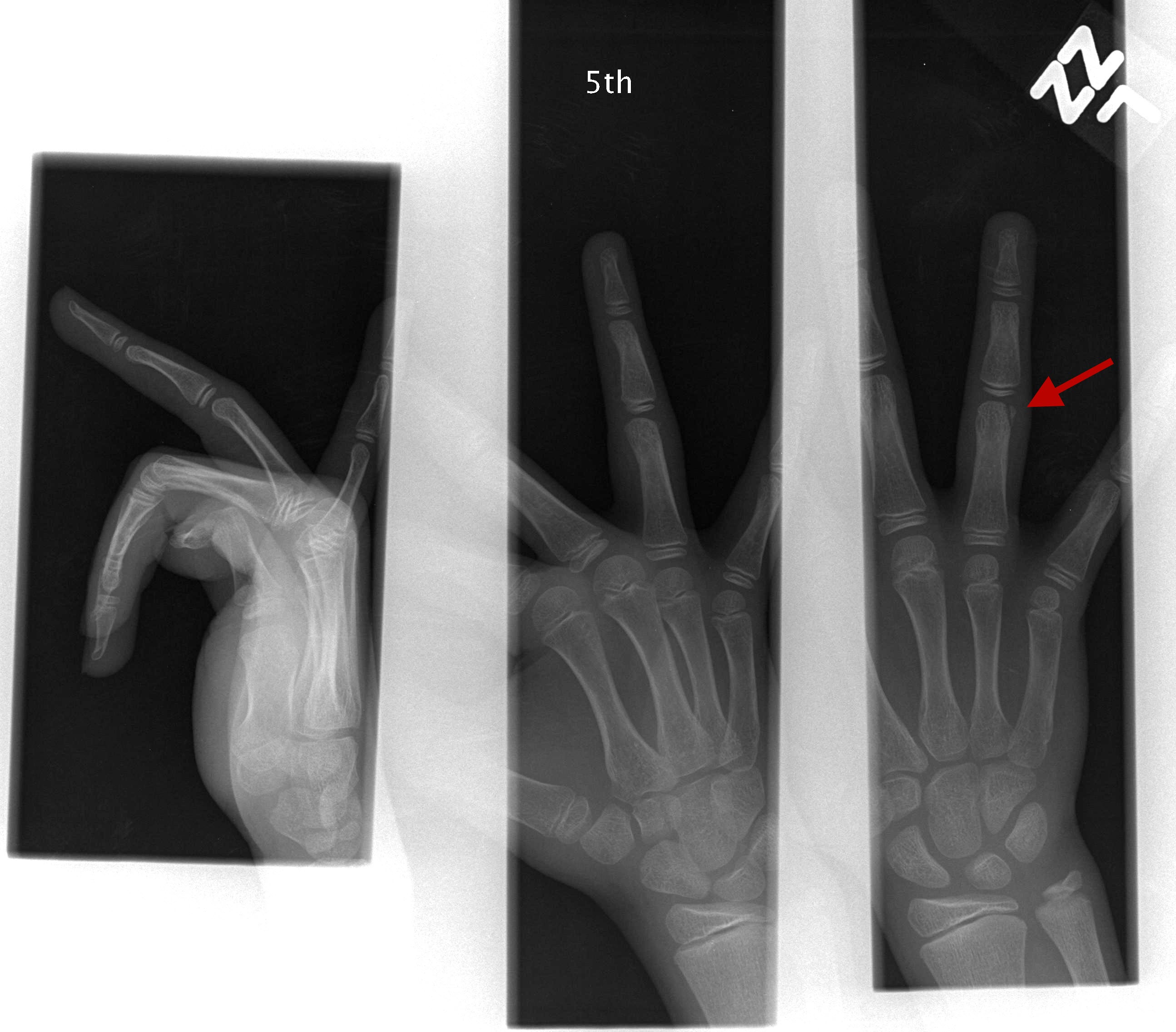
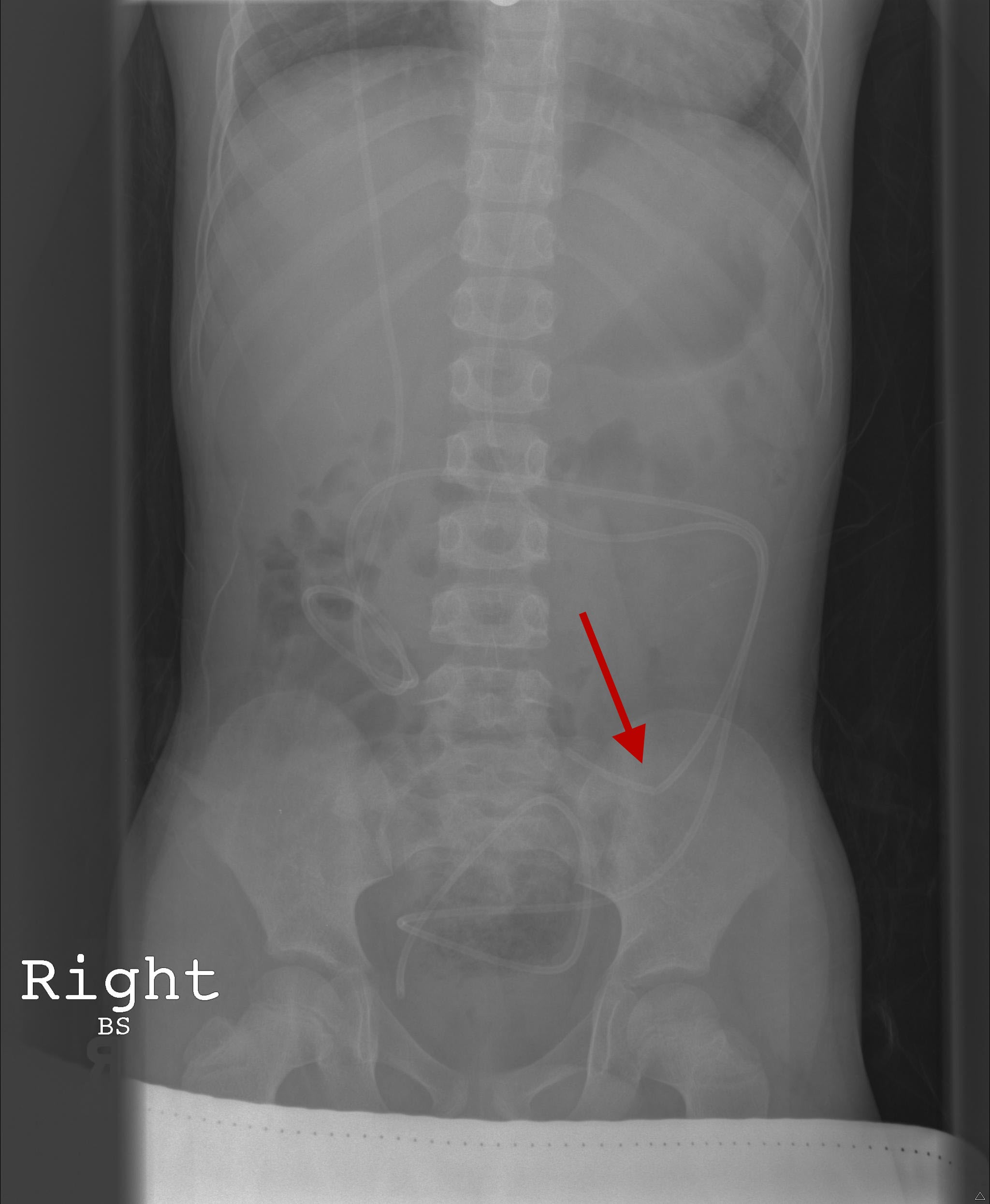
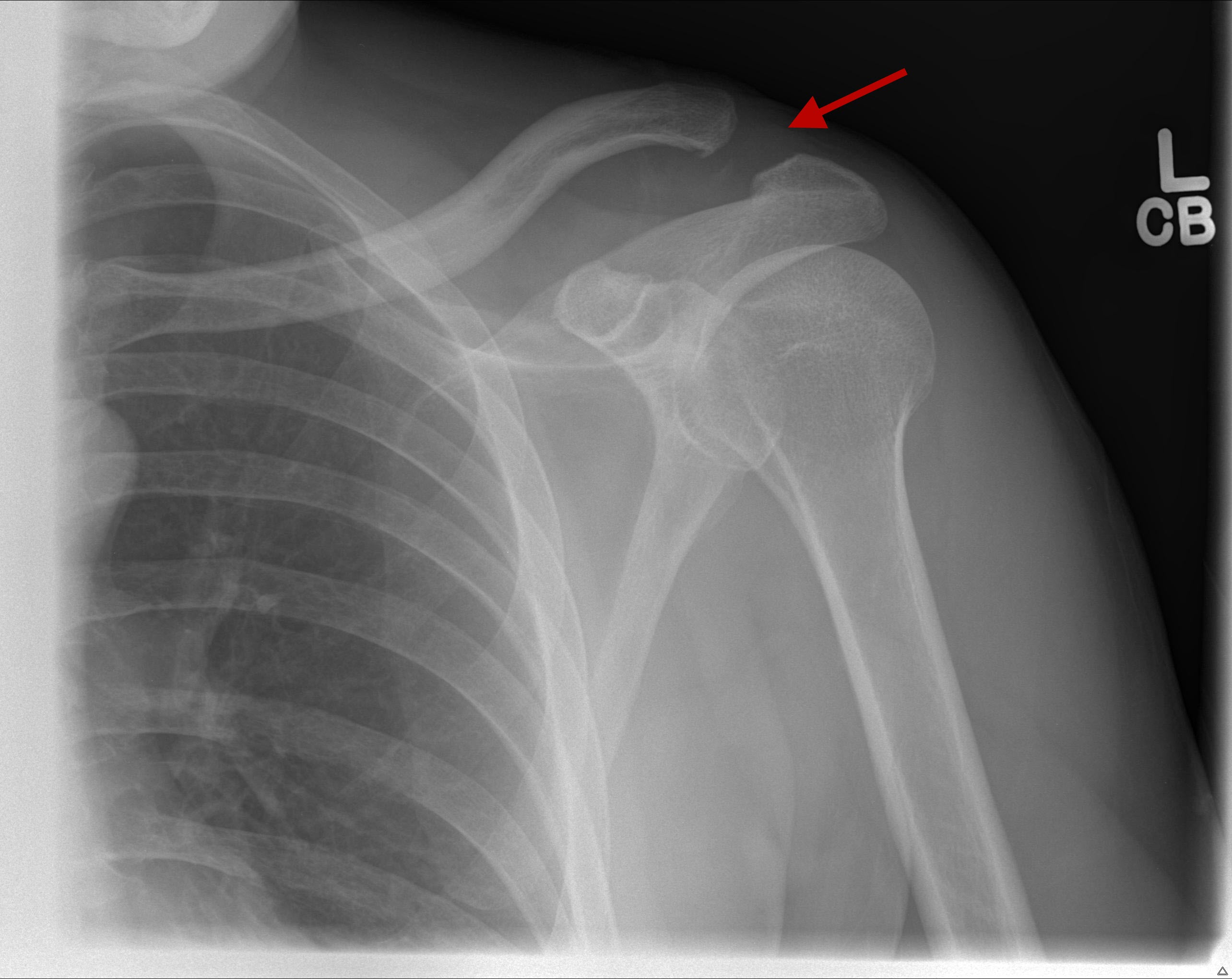
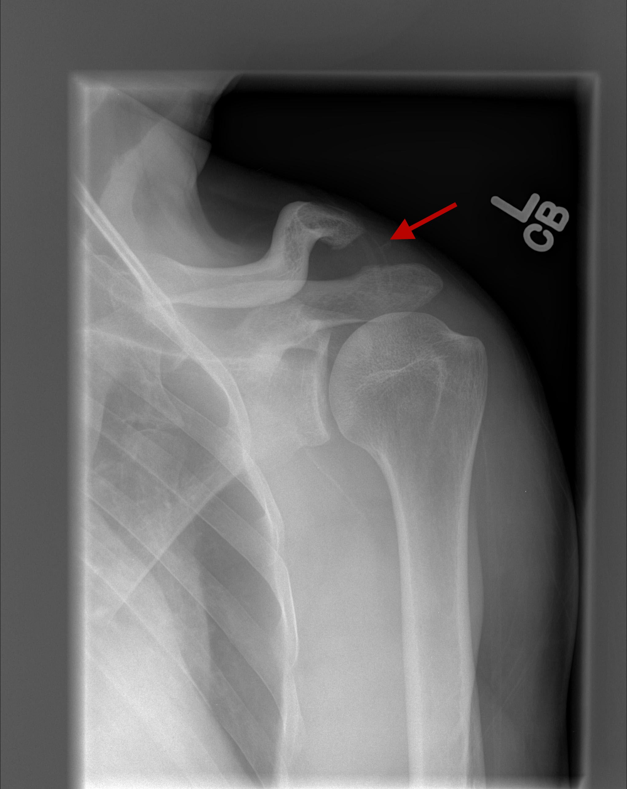
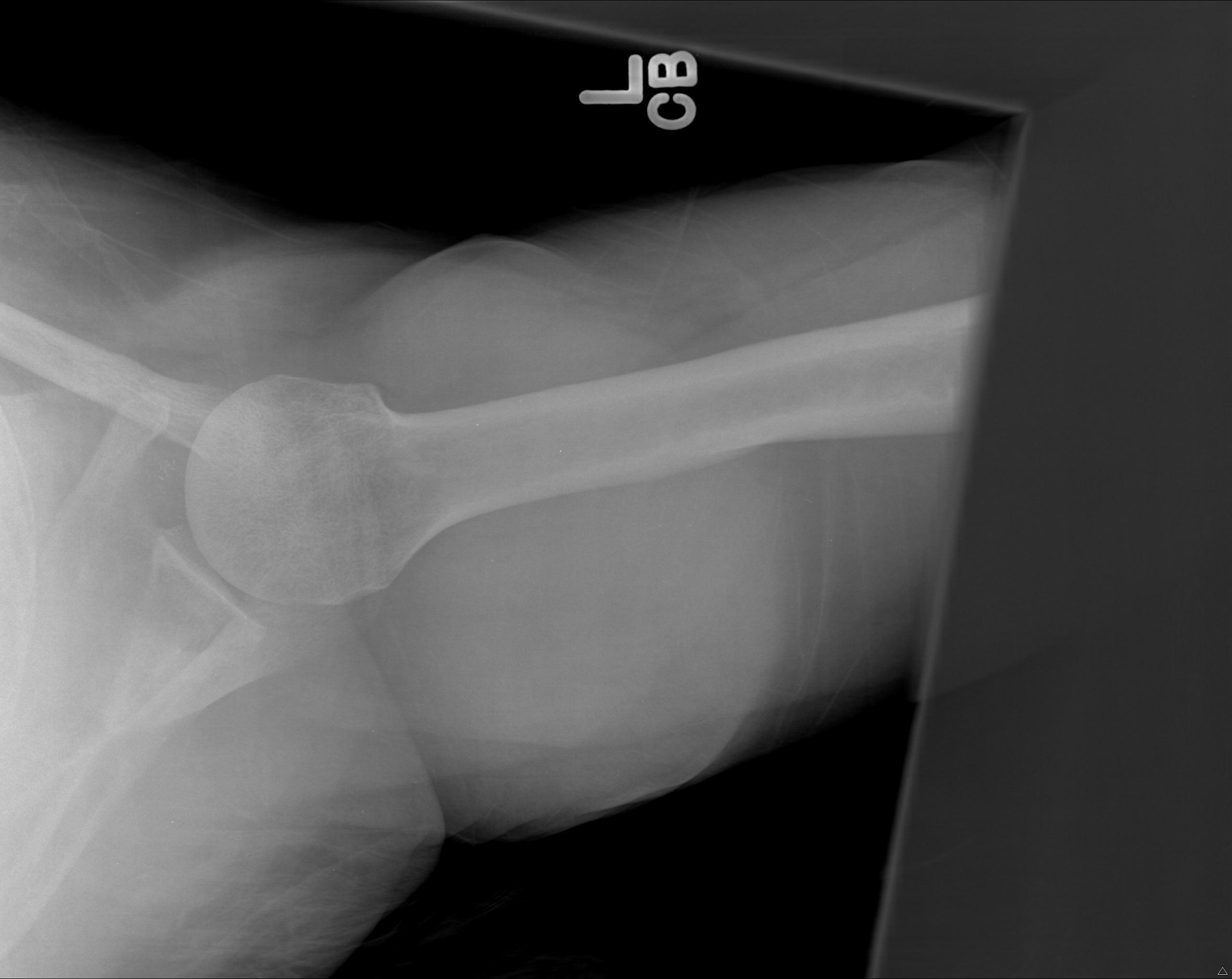
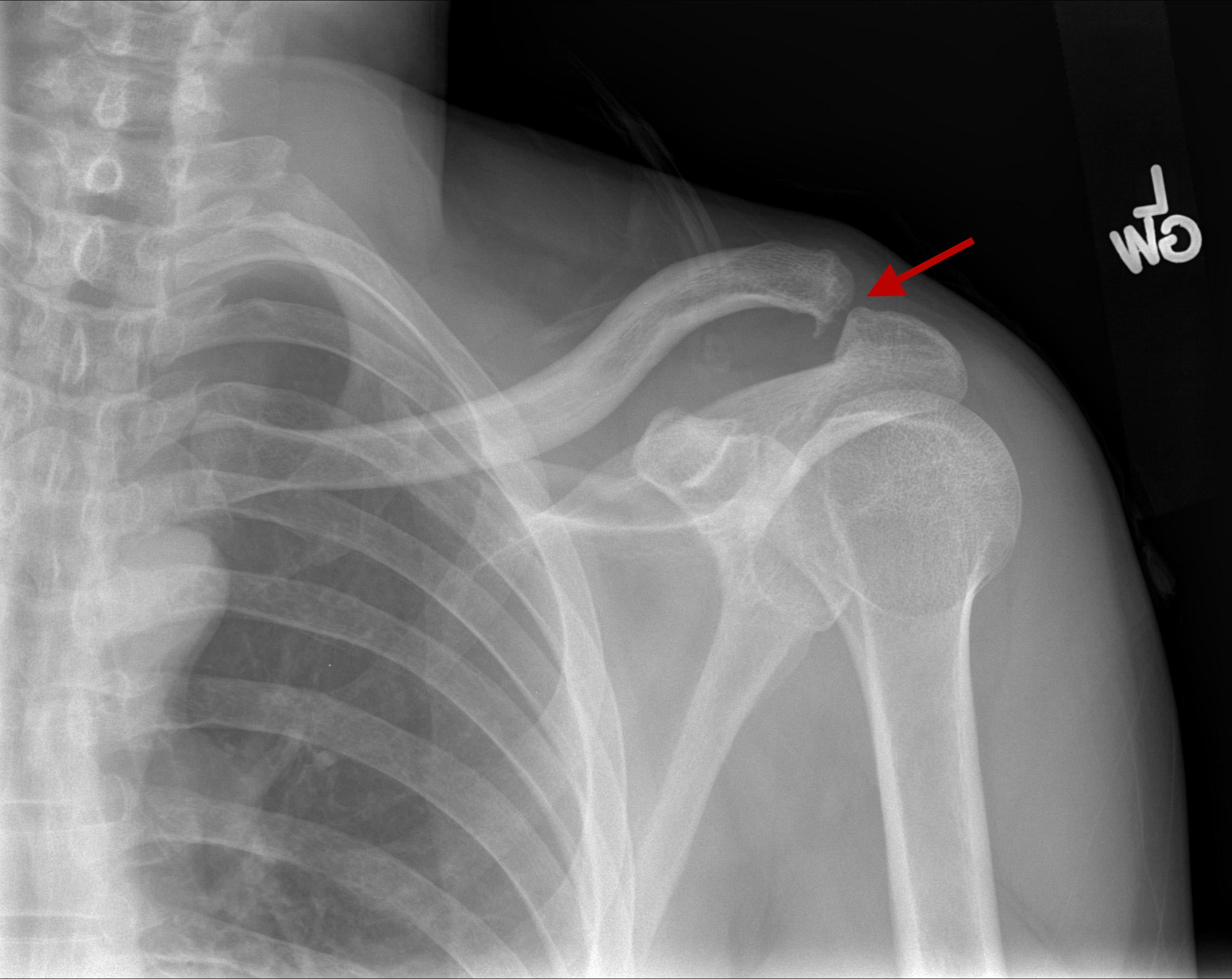
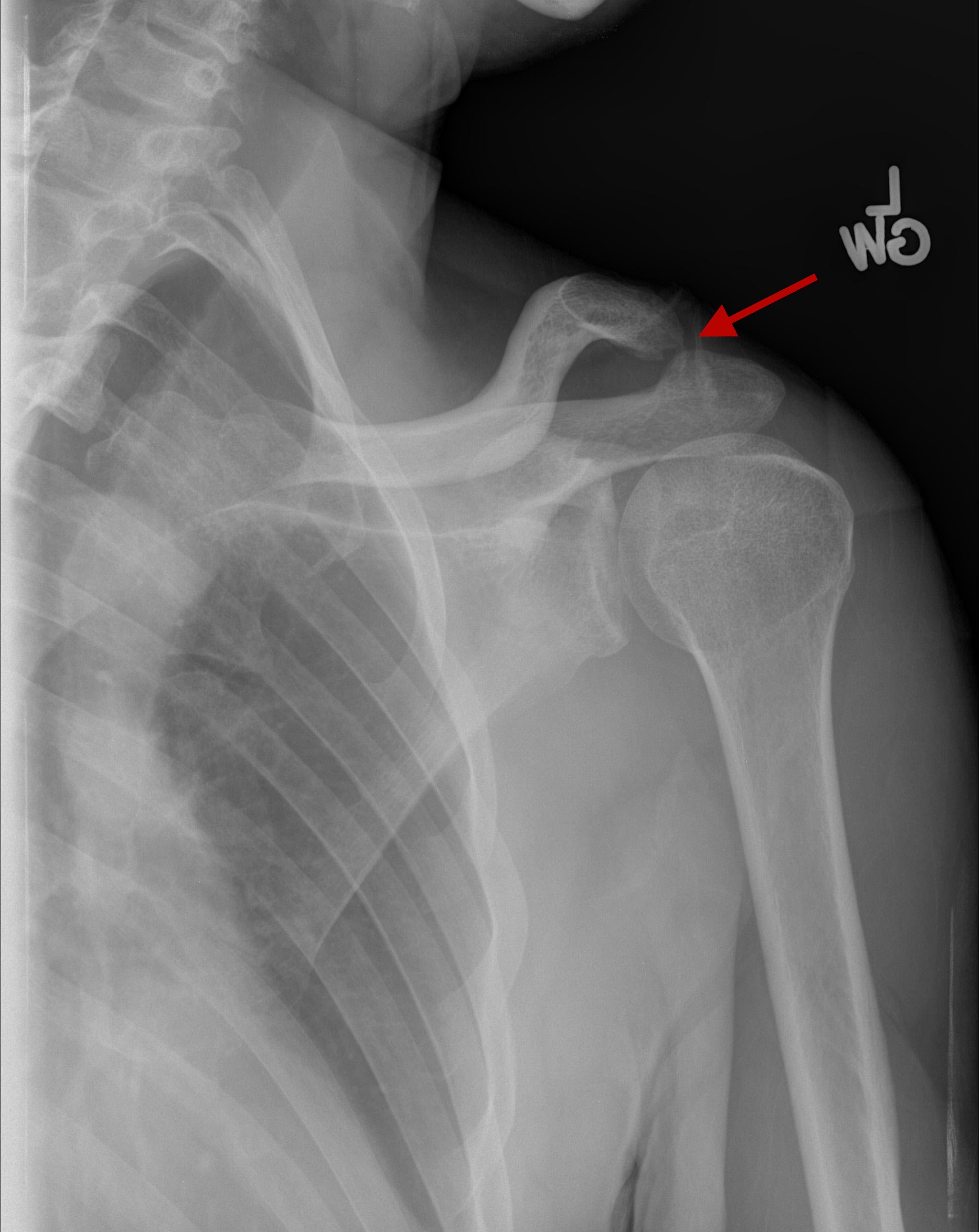
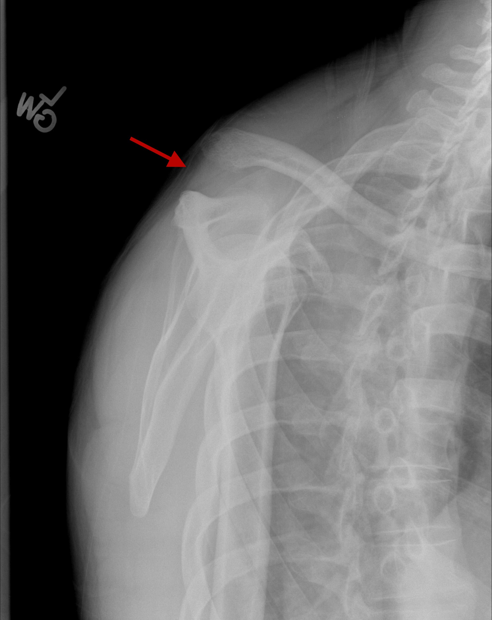
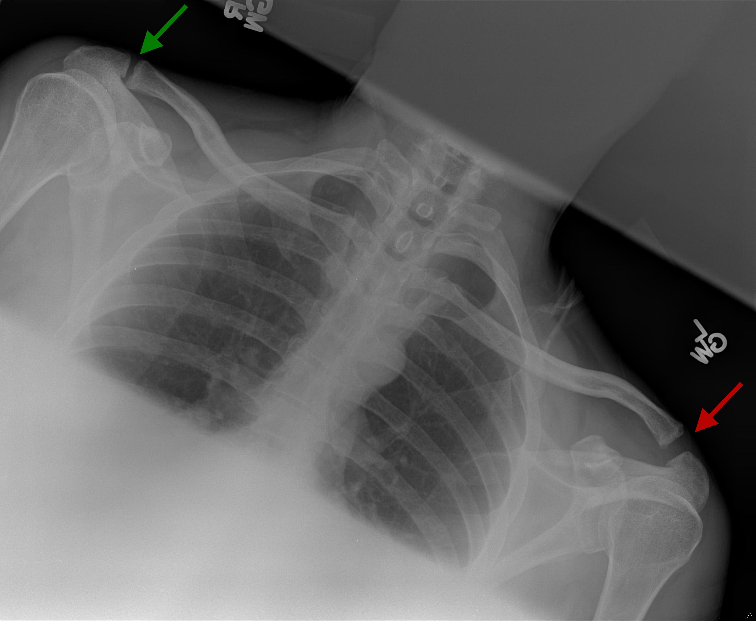
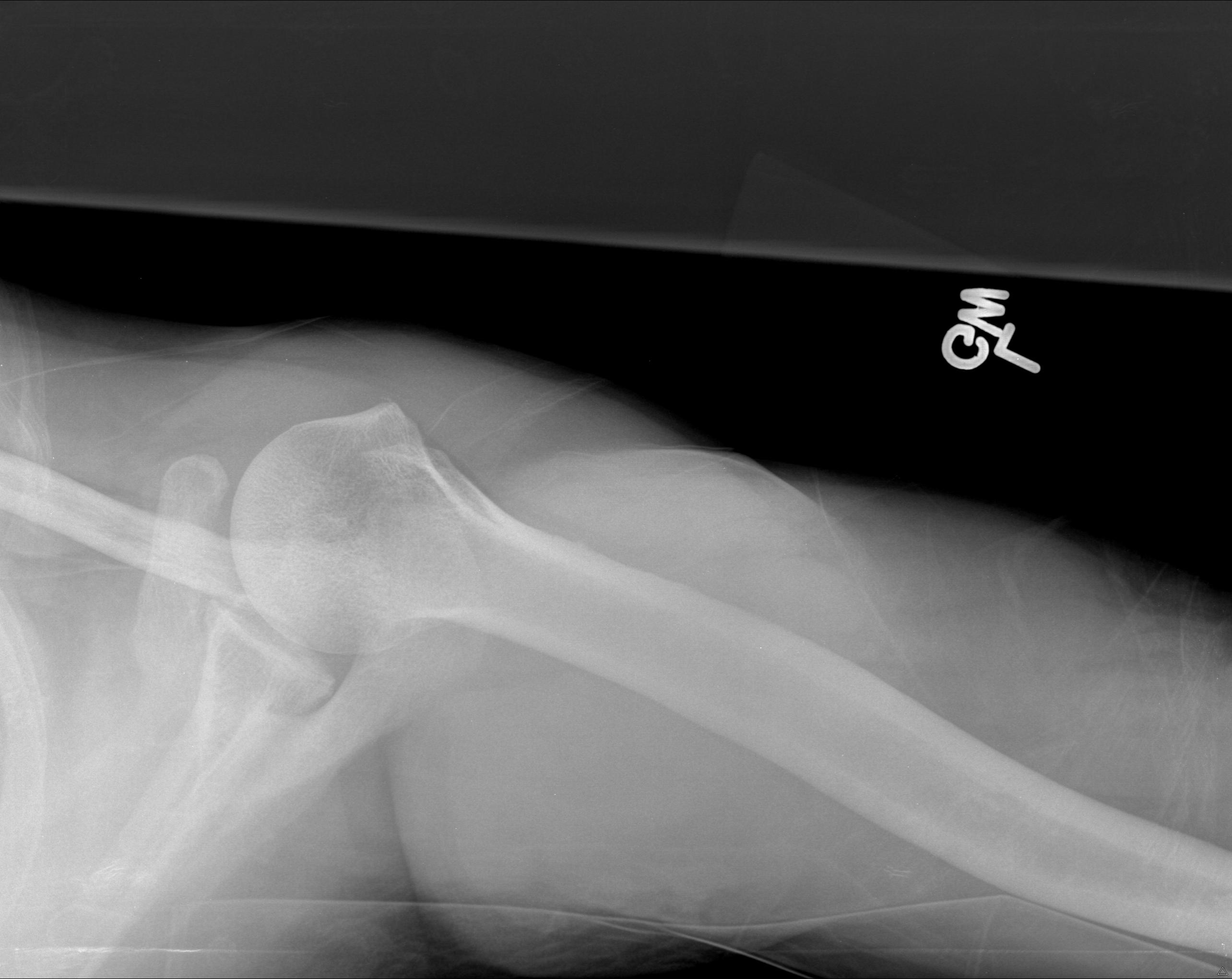
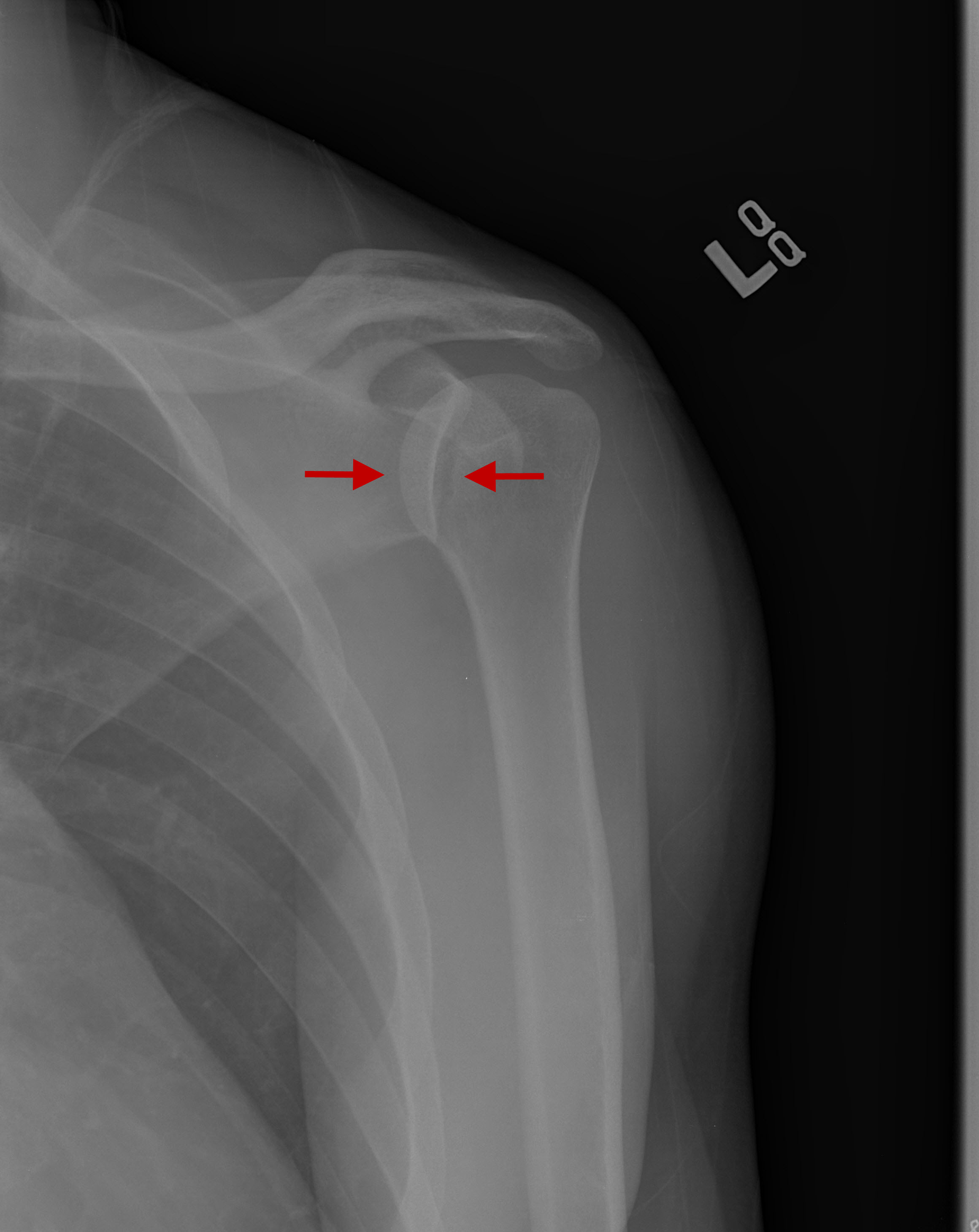
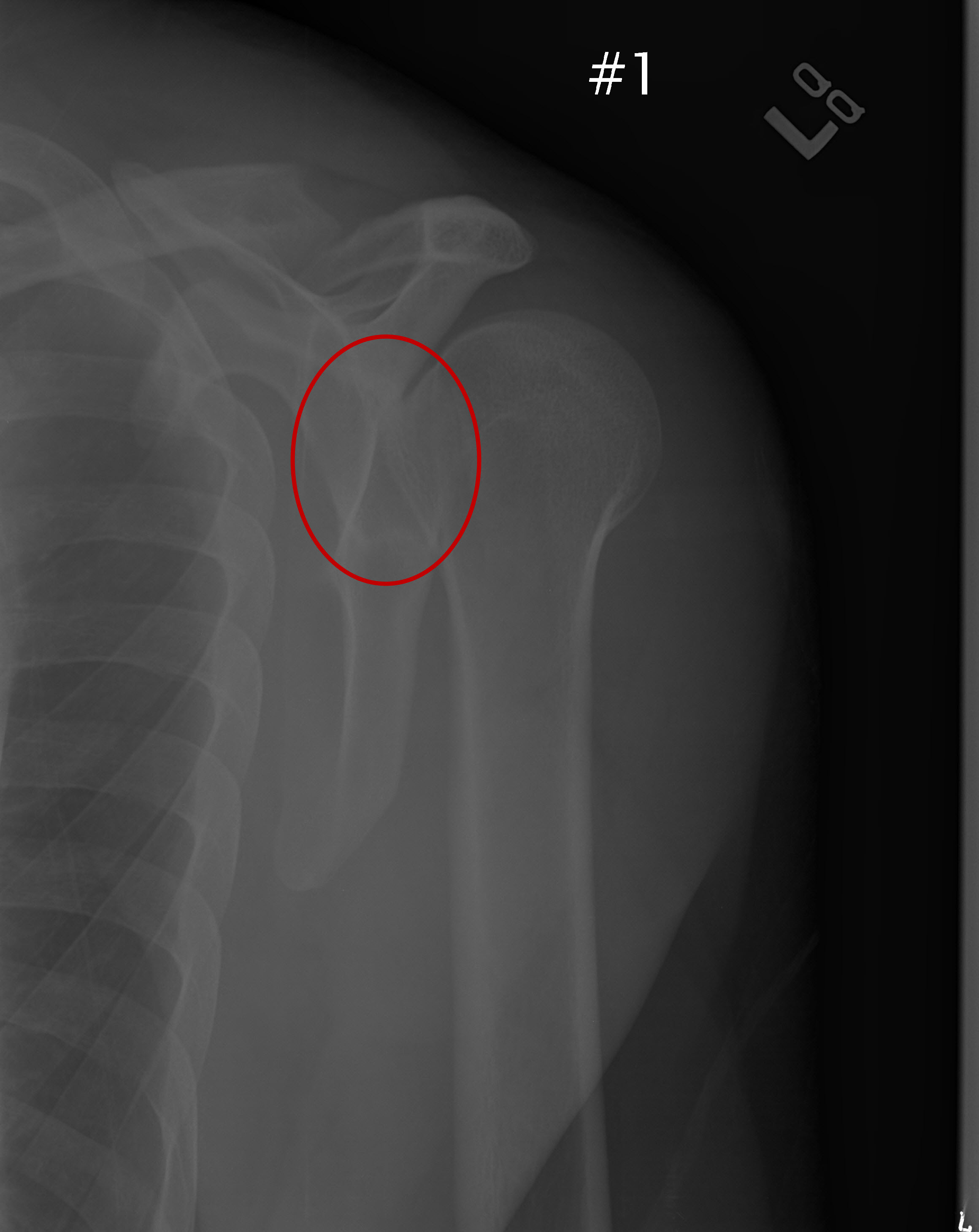
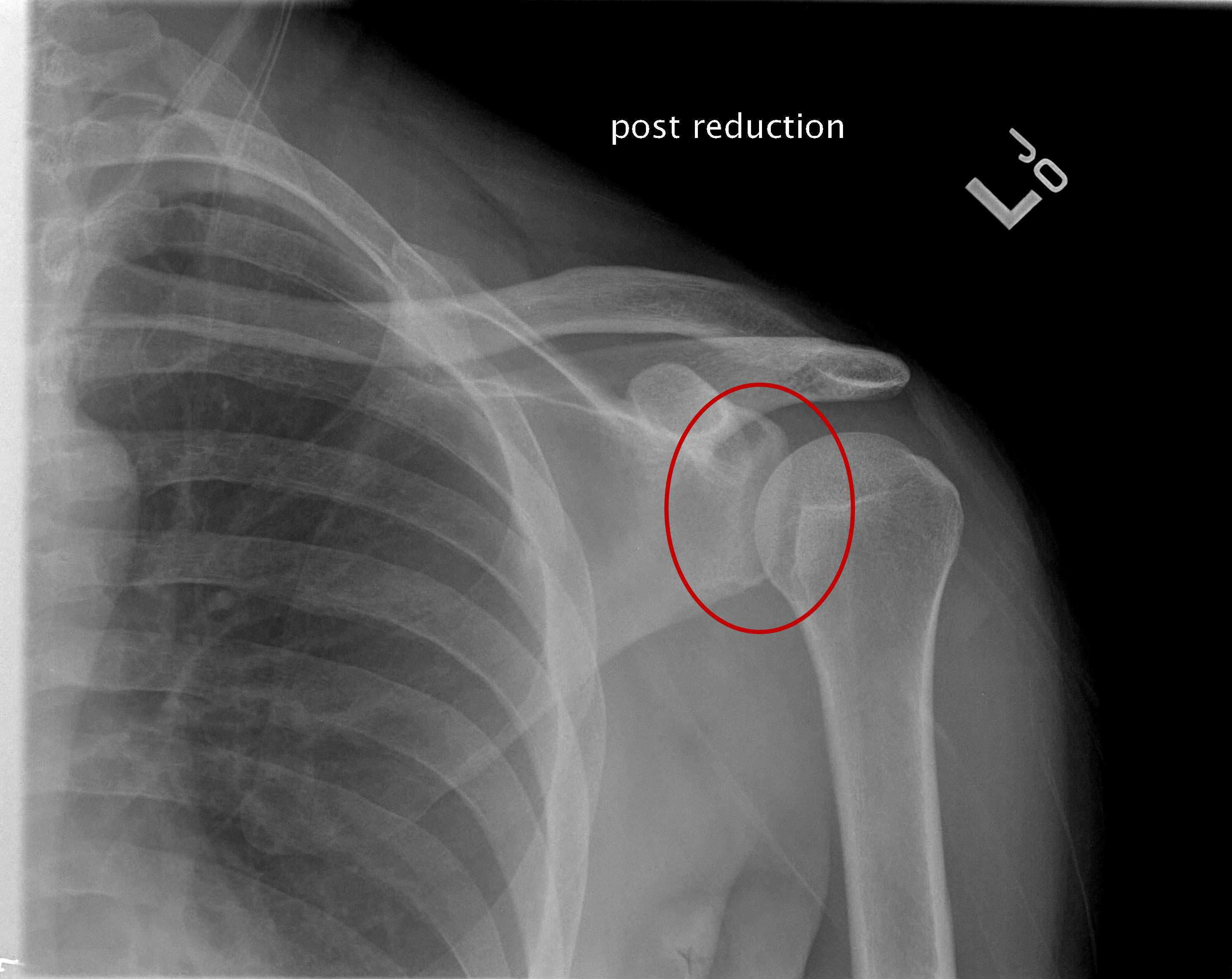
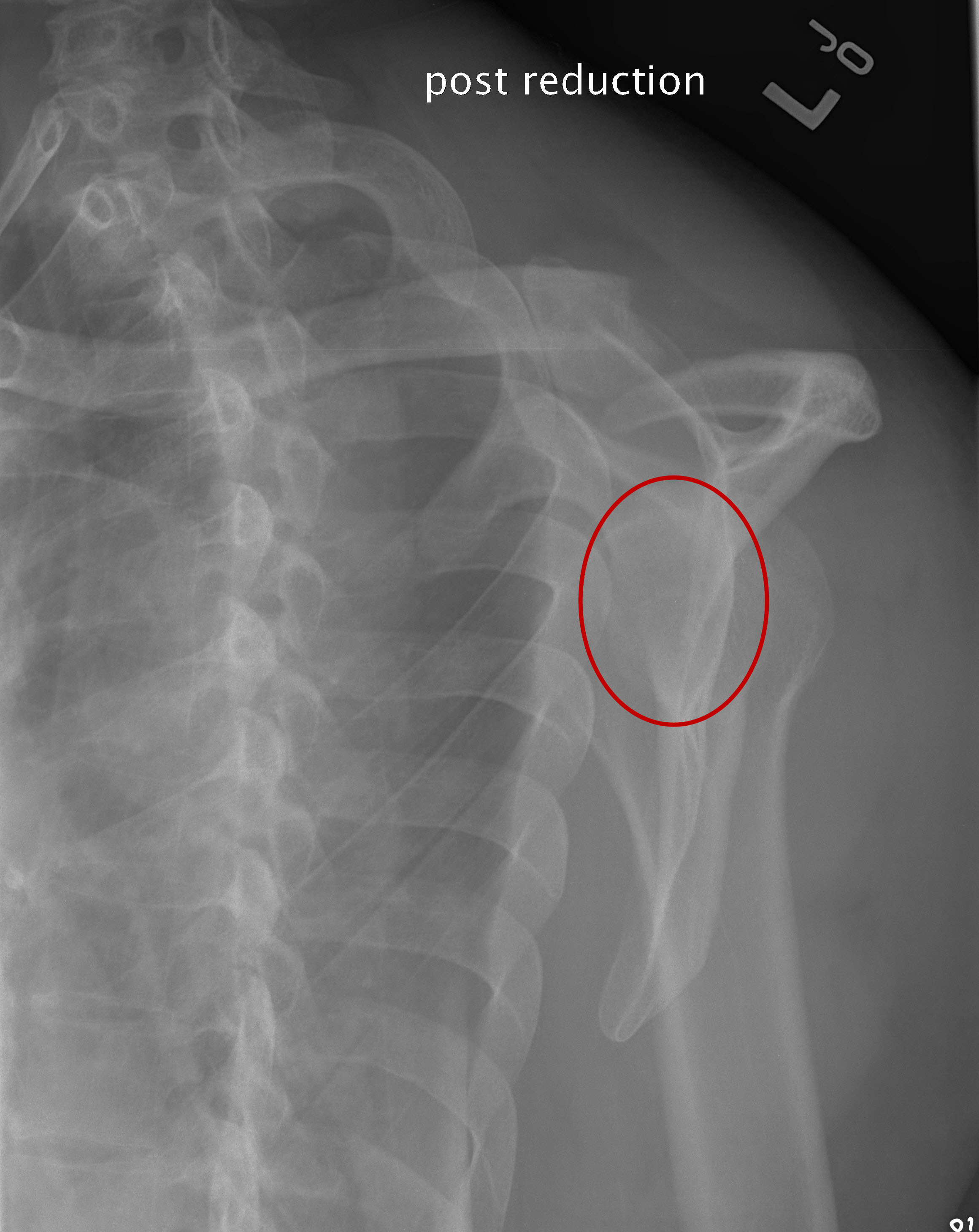
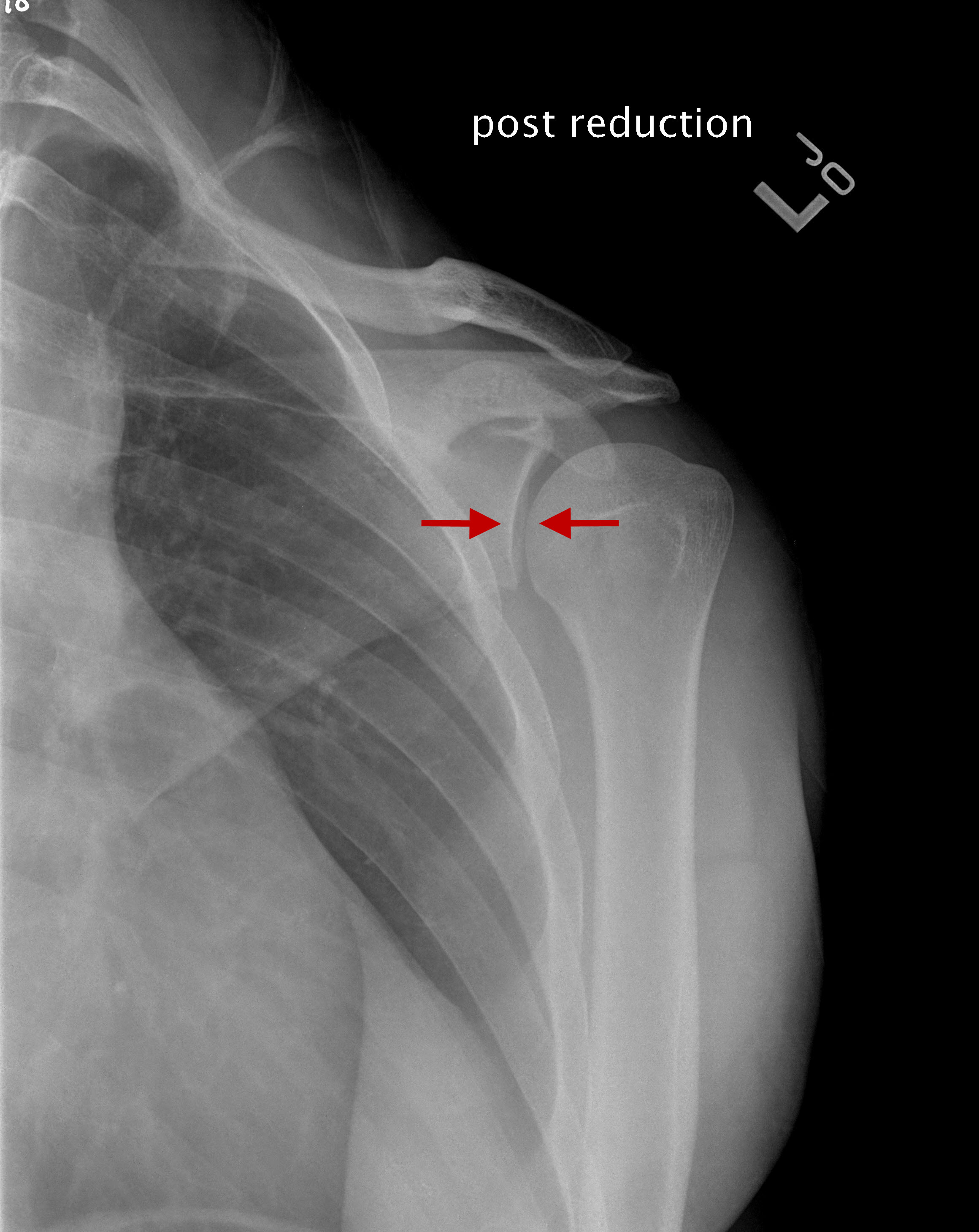
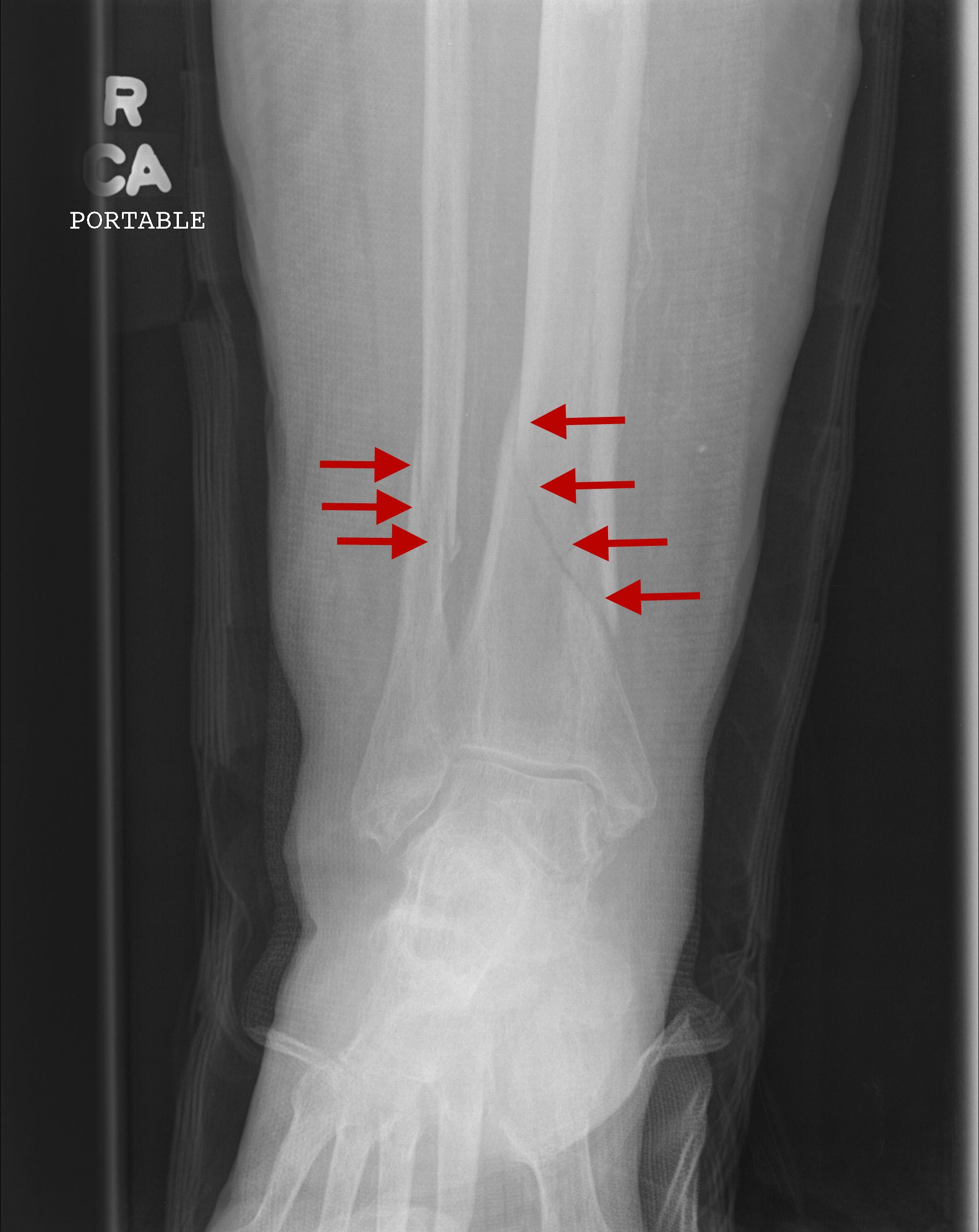
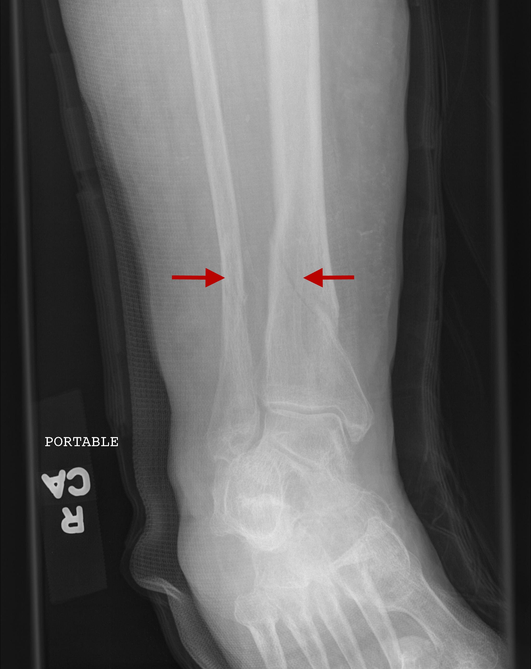
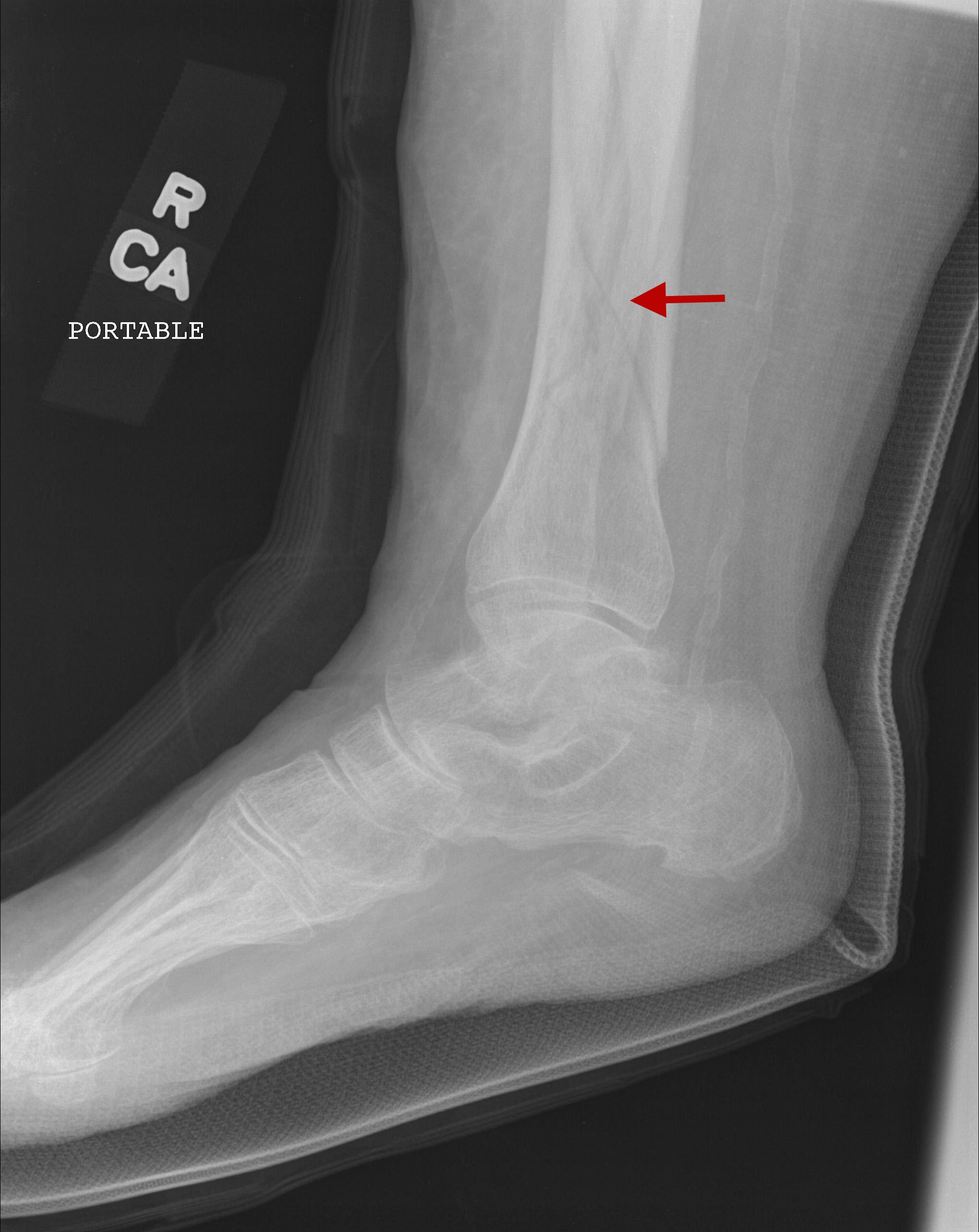
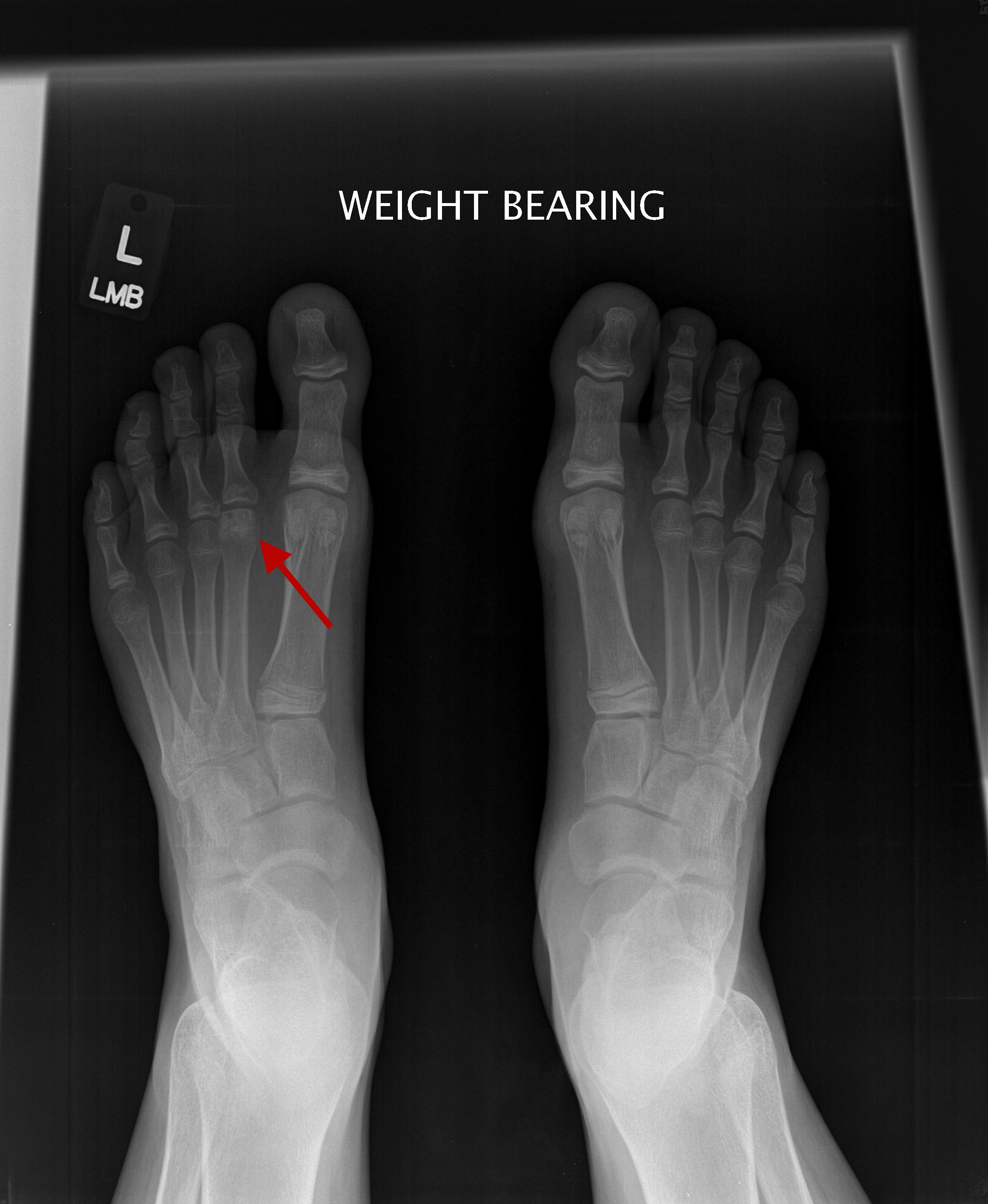
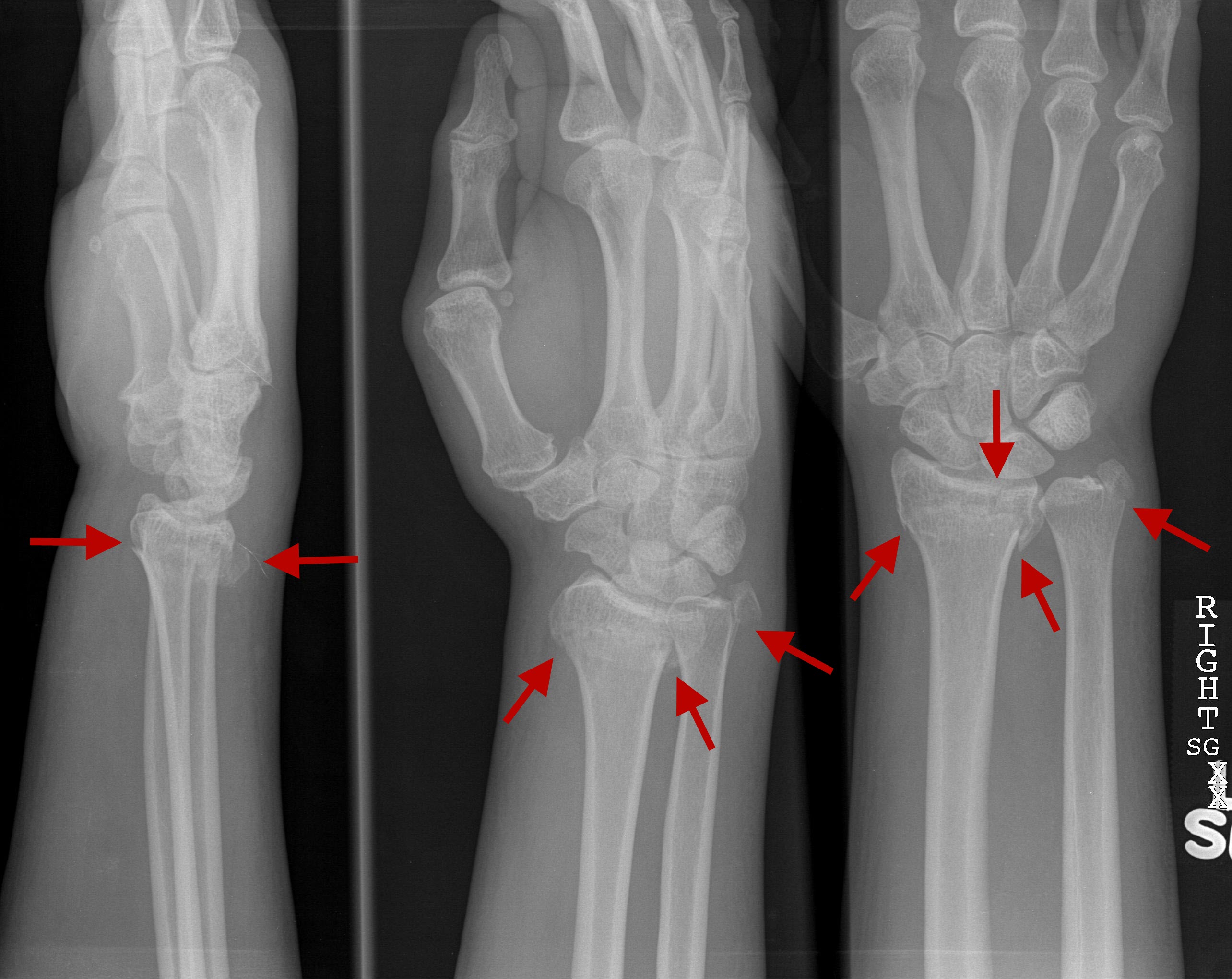
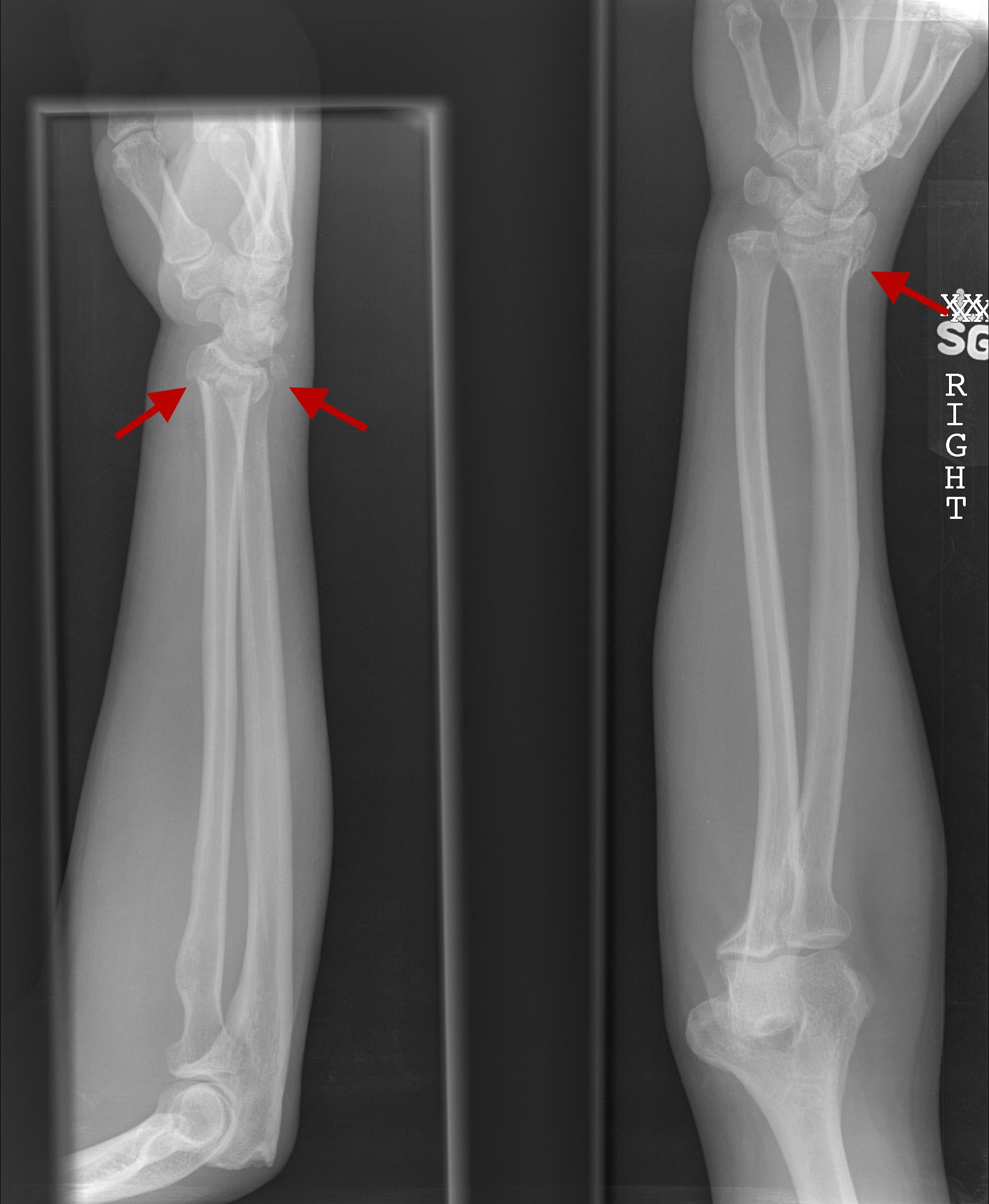
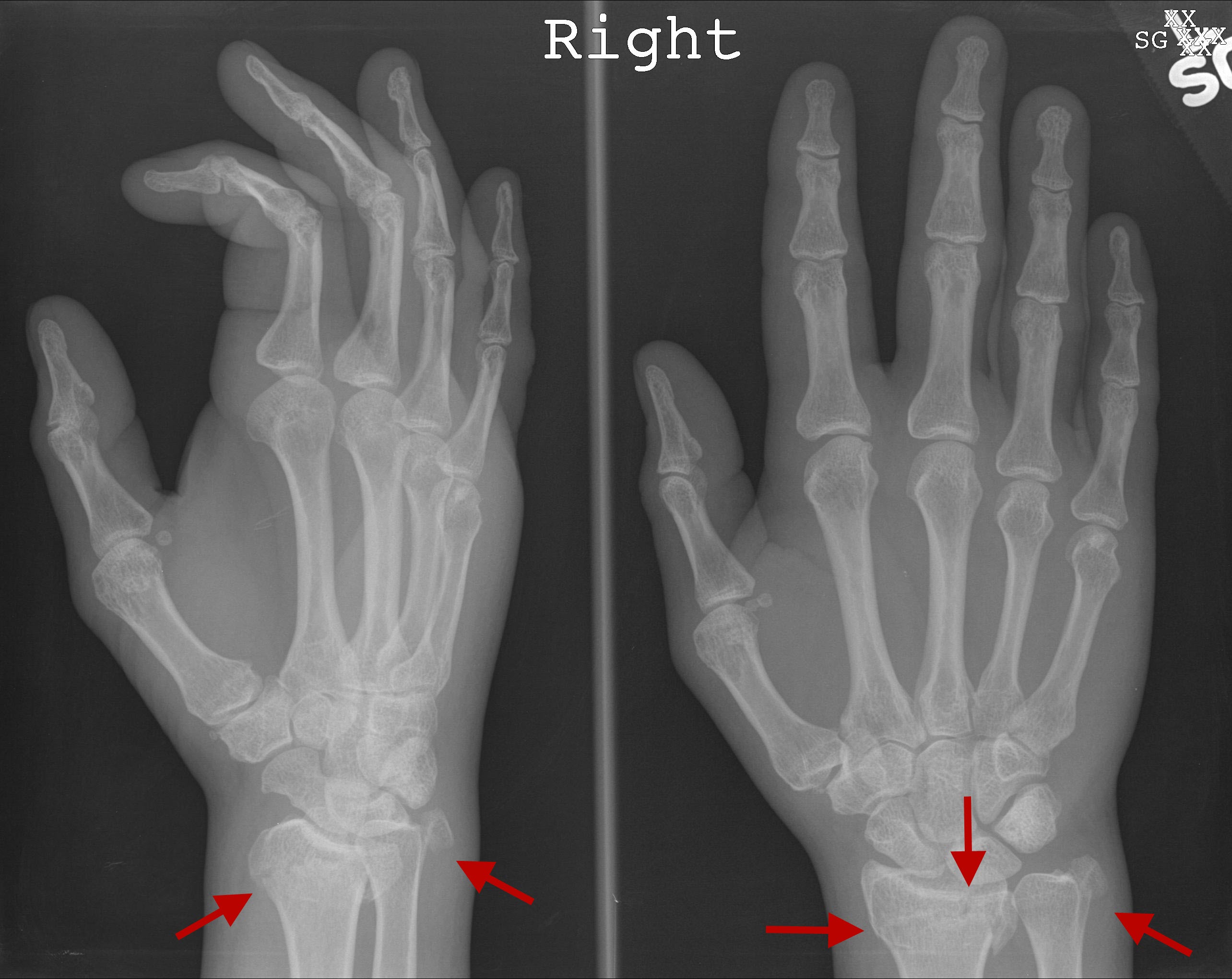
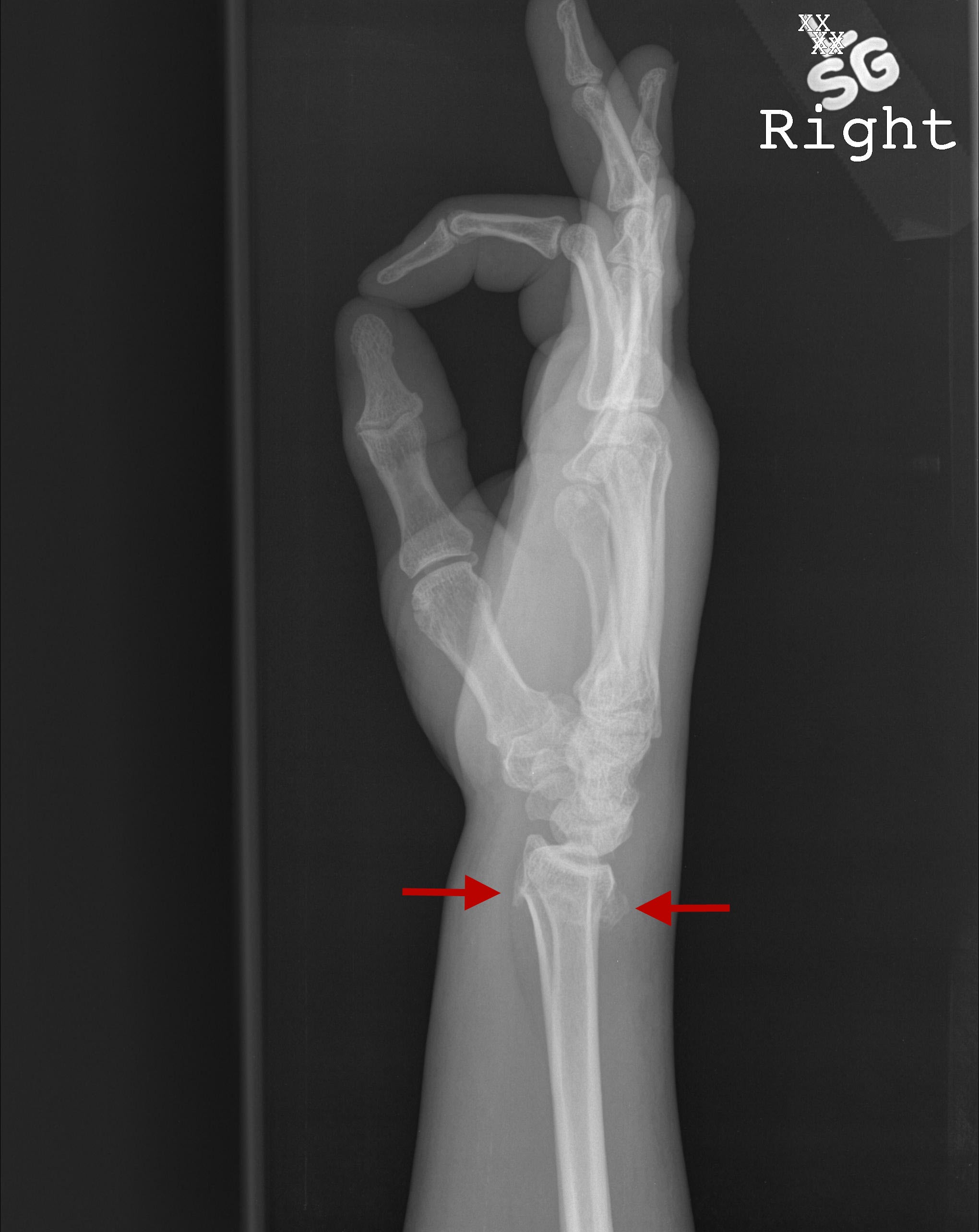
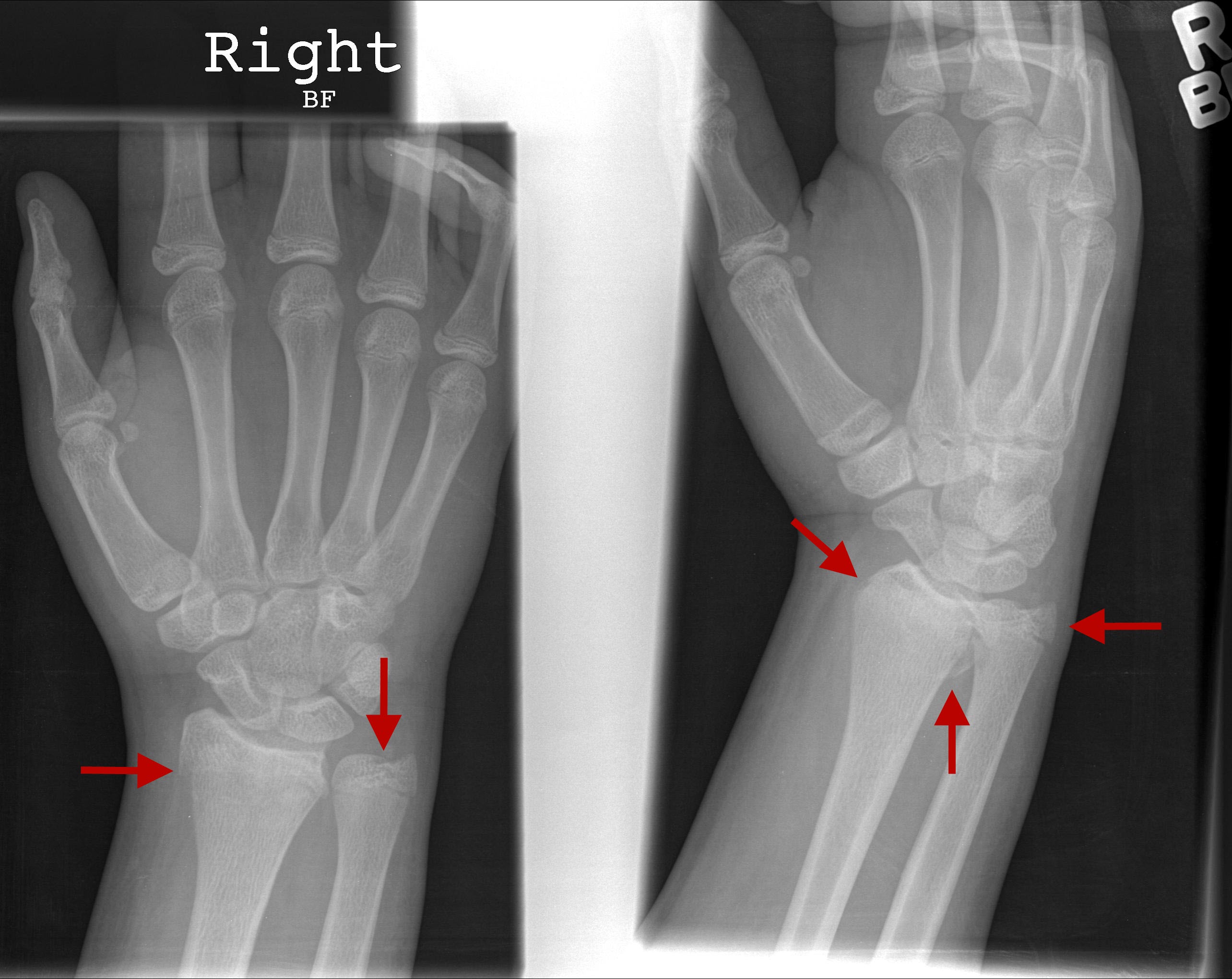
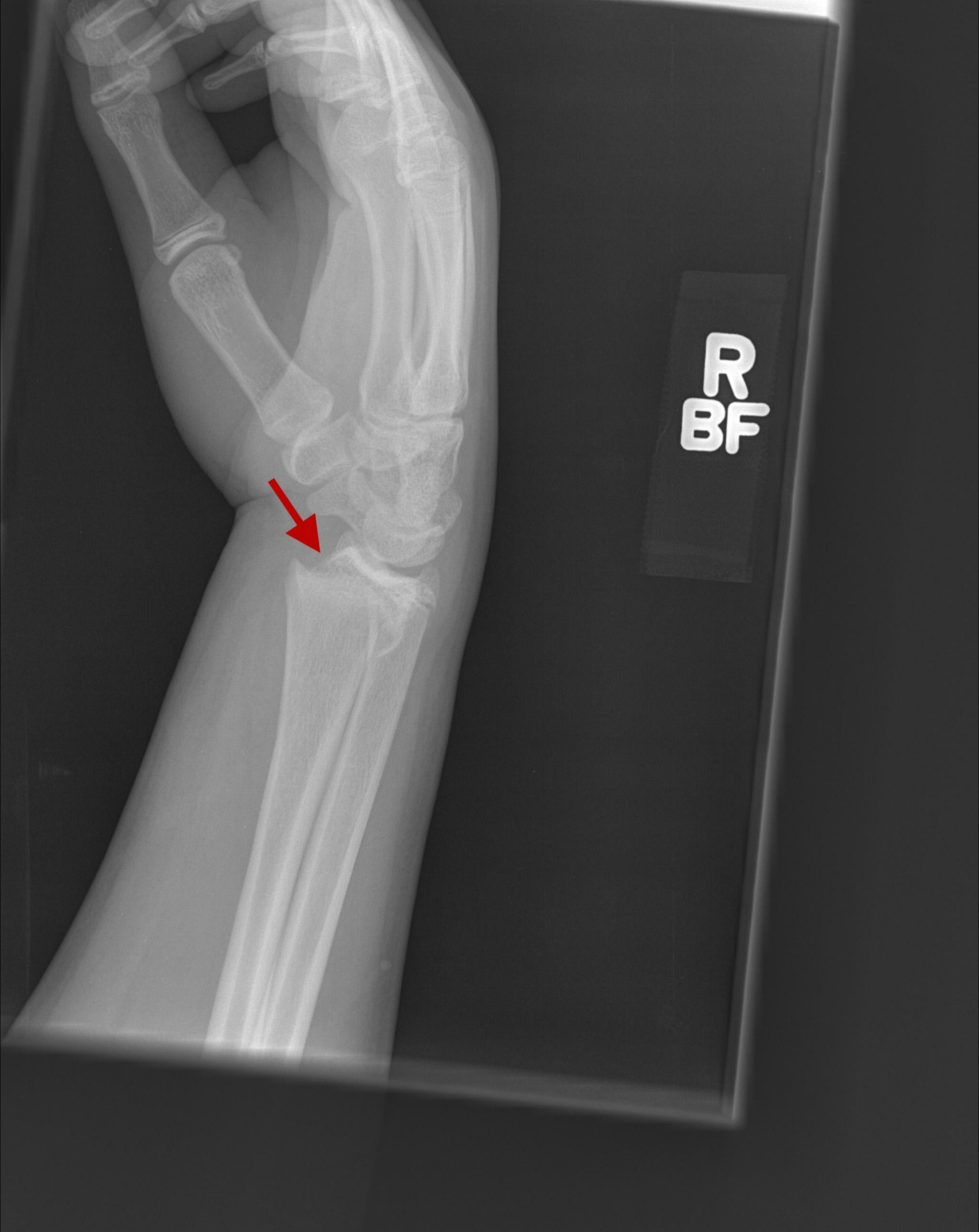
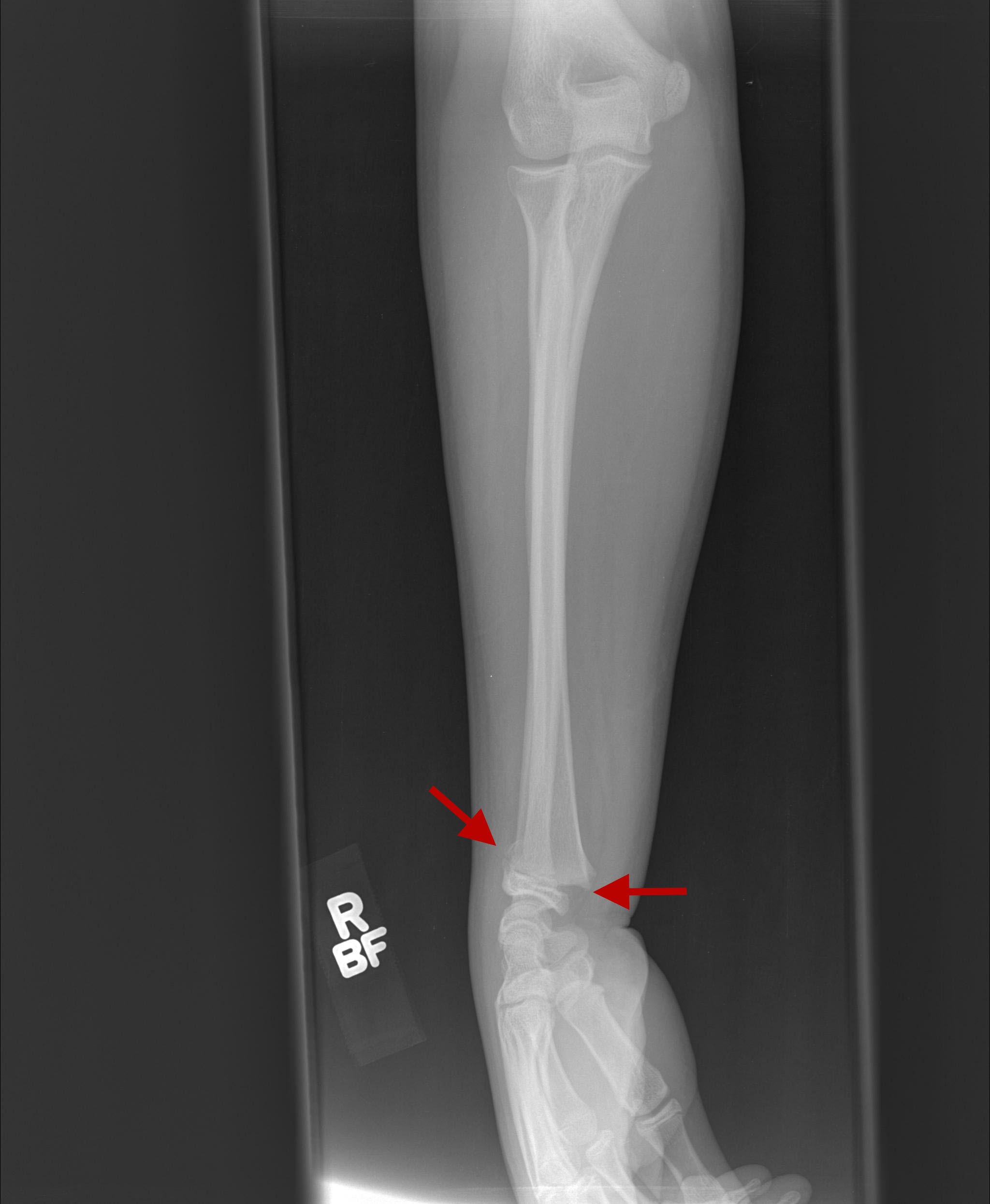
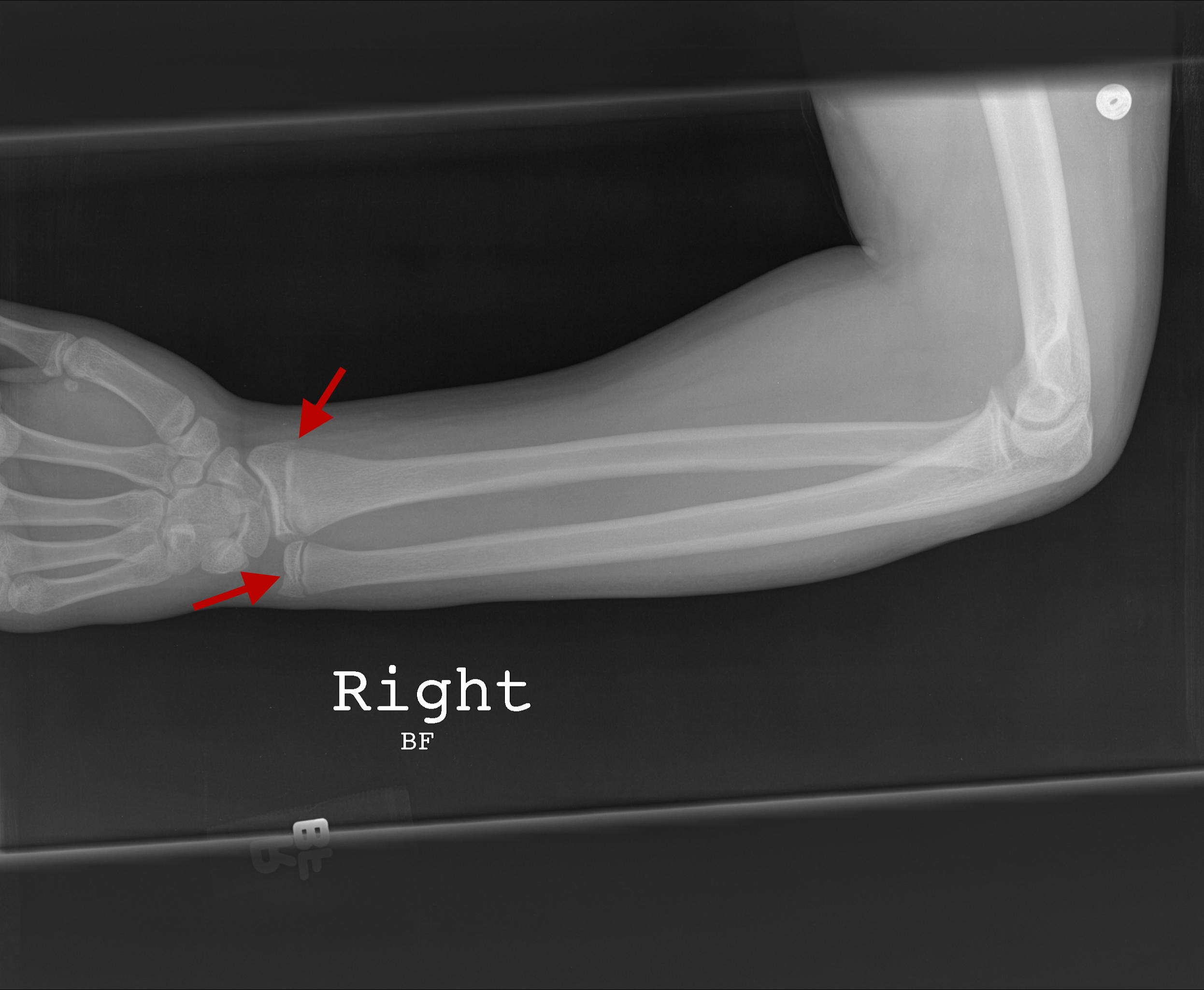
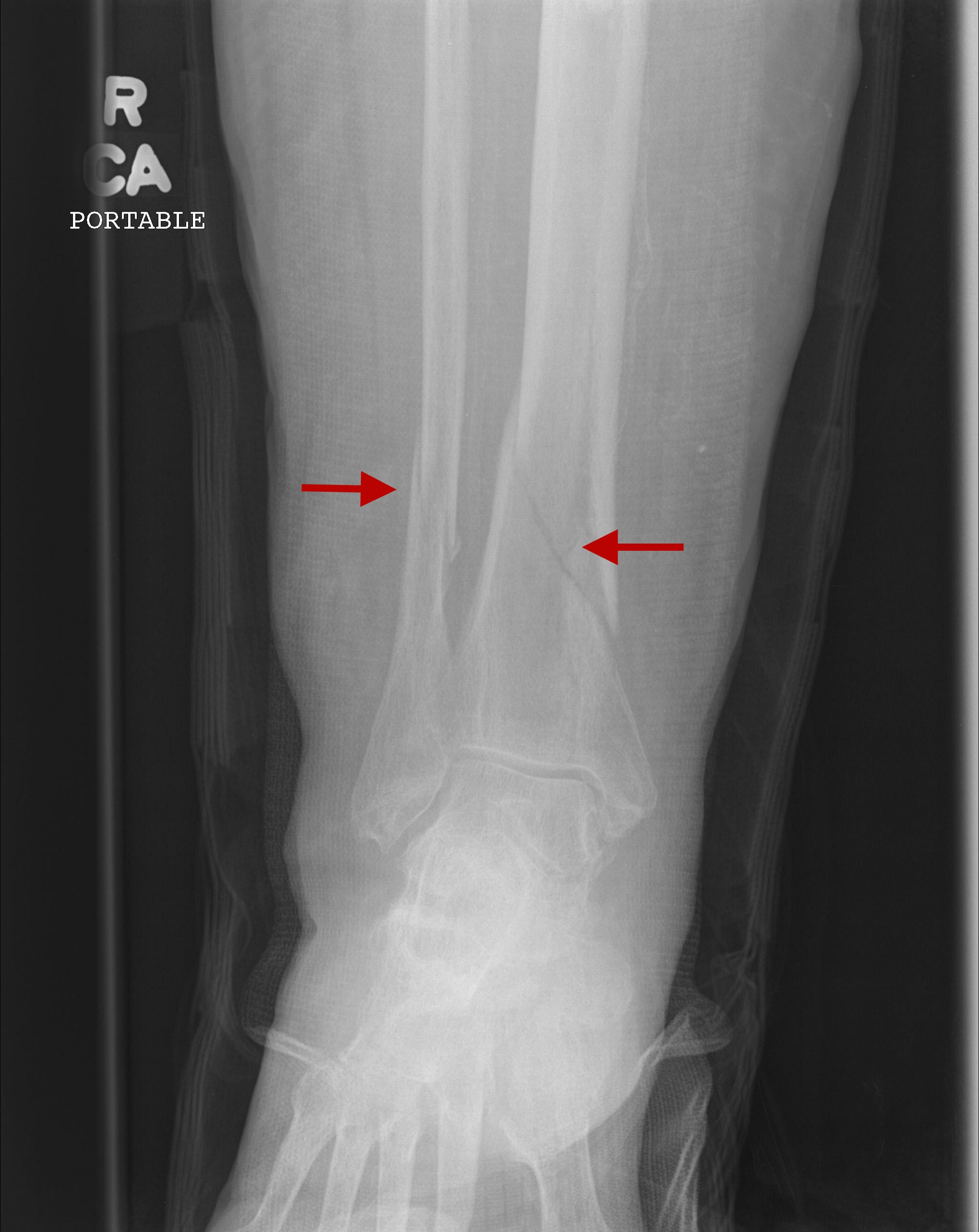
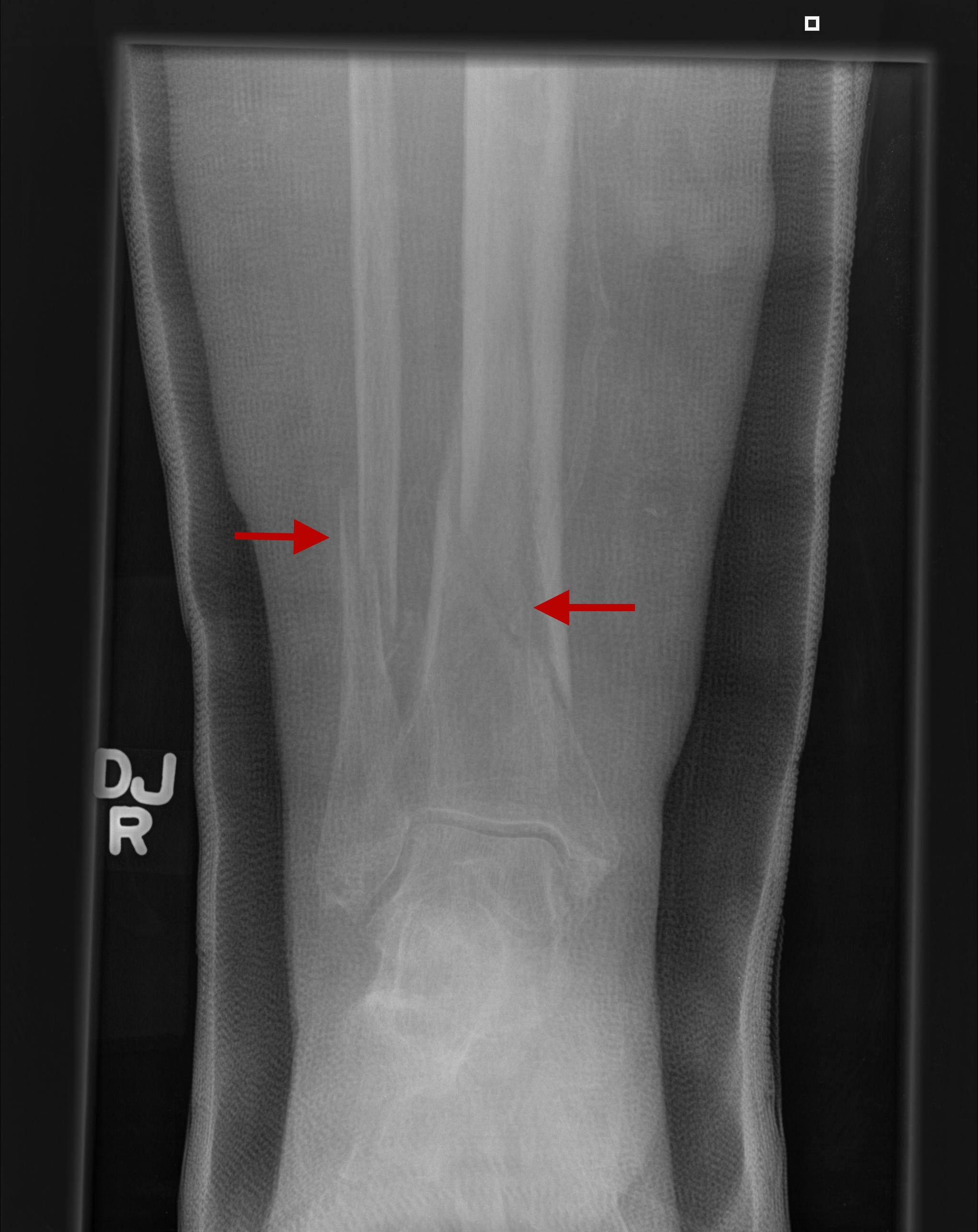
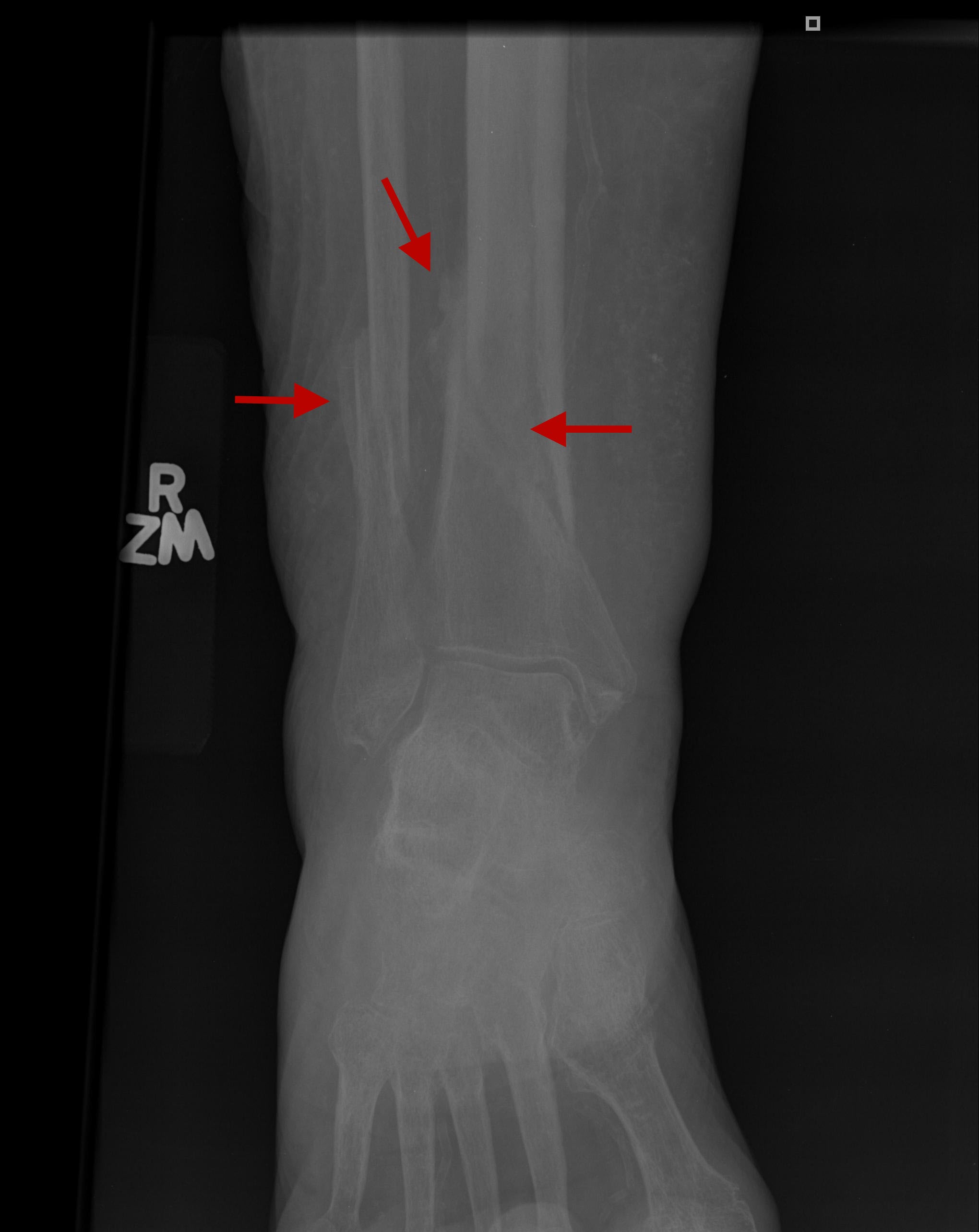
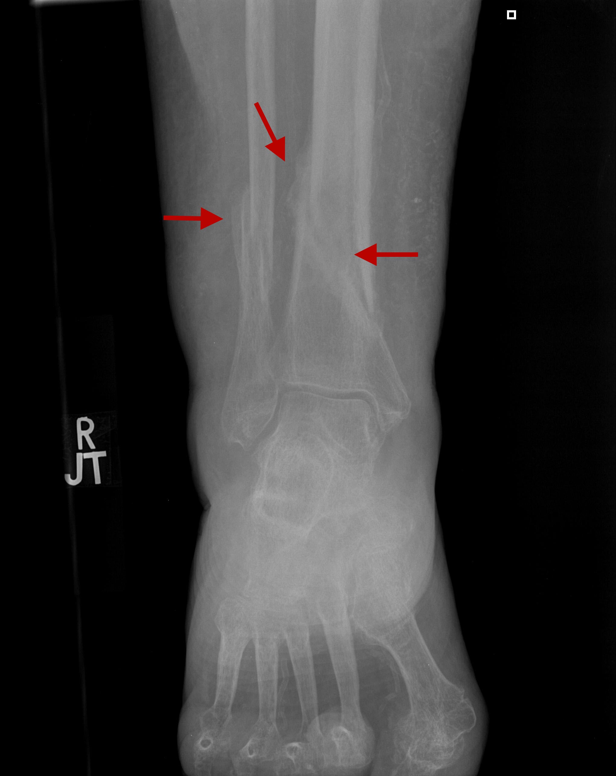
MSKDislocations - Dislocated left hip prosthesis Trauma - Comminuted intertrochanteric fracture Trauma - Buckle fracture base of 4th phalanx Trauma - Clavicular Bayonette fracture Trauma - Patellar tendon rupture Trauma - Intraarticular corner fracture Devices - Kinked VP shunt Trauma - Acromioclavicular ligament tear Trauma - Posterior shoulder dislocation Trauma - Distal tibial and fibular fracture Vascular - Freiberg infraction Trauma - Distal radius and ulnar styloid fracture Trauma - Distal radius and ulnar head fracture Trauma - Fracture healing process MSK: FracturesDiagnosis: Distal fibular fracture (Plain film, Radiograph)There is a complete spiral fracture of the distal fibula. Three is also associated soft tissue swelling around the ankle (green arrows).MSK: DislocationsDiagnosis: Dislocated left hip prosthesis (Plain film, Radiograph)The left hip prosthesis has dislocated superiorly and anteriorly.MSK: TraumaDiagnosis: Comminuted intertrochanteric fracture (Plain film, Radiograph)There is a comminuted intratrochanteric fracture of the right hip with avulsion of the lesser and greater trochanters, superior migration, and moderate varus angulation. The crosstable lateral examination demonstrates mild posterior angulation.MSK: TraumaDiagnosis: Buckle fracture base of 4th phalanx (Plain film, Radiograph)There is a buckle fracture at the ulnar aspect of the base of the fourth proximal phalanx. There is surrounding soft tissue swelling.MSK: TraumaDiagnosis: Clavicular Bayonette fracture (Plain film, Radiograph)Transverse fracture approximately 3 cm medial to the distal end of the clavicle in bayonet appositionMSK: TraumaDiagnosis: Patellar tendon rupture (Plain film, Radiograph)Patella alta (high riding patella in relation to femur), joint effusion and soft tissue swellingMSK: TraumaDiagnosis: Intraarticular corner fracture (Plain film, Radiograph)There is a minimally displaced intraarticular oblique corner fracture of the fourth proximal phalanx head on the ulnar side. There is minimal associated soft tissue swelling. There is no other fracture or dislocation. No periosteal reaction or cortical thickening is present.MSK: DevicesDiagnosis: Kinked VP shunt (Plain film, Radiograph)The distal end of the second shunt catheter is kinkedMSK: TraumaDiagnosis: Acromioclavicular ligament tear (Plain film, Radiograph)Images 1-3 demonstrate widening of the AC interval, compatible with AC ligament tear. Images 4-8 are comparison images obtained one year earlier, demonstrating mild widening of the AC interval, however noyt in such extend.MSK: TraumaDiagnosis: Posterior shoulder dislocation (Plain film, Radiograph)Images 1-2 demonstrate an empty glenoid sign with the humeral head in malposition in relation to the glenoid. Images 3-5 are post reduction, demonstrating the humeral head in anatomic position with the glenoid.MSK: TraumaDiagnosis: Distal tibial and fibular fracture (Plain film, Radiograph)There are oblique complete fractures of the distal tibia and fibula.MSK: VascularDiagnosis: Freiberg infraction (Plain film, Radiograph)There is flattening and sclerosis of the second metatarsal head, compatible with Freiberg infraction.MSK: TraumaDiagnosis: Distal radius and ulnar styloid fracture (Plain film, Radiograph)There is a comminuted fracture of the distal radius as well as the ulnar styloid.MSK: TraumaDiagnosis: Distal radius and ulnar head fracture (Plain film, Radiograph)There is a Salter-Harris II fracture of the distal radius and Salter-Harris III fracture of the distal ulna.MSK: TraumaDiagnosis: Fracture healing process (Plain film, Radiograph)Seen is chronologic healing progress of a distal tibial and fibular fracture. Image 1 is at the time of fracture. Image 2 is 3 weeks later, image 3 is 6 weeks later, image 4 is 8 weeks later and image 5 is 15 weeks later. Nnote the progression of callus formation.Home :: Contact Us Make On Call Radiology your homepage :: Add On Call Radiology to your favorites |